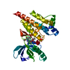
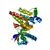
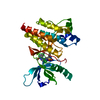
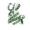
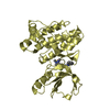
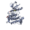
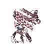
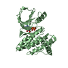
Entry Database : PDB / ID : 2ijmTitle Crystal Structure of Focal Adhesion Kinase Domain with 2 molecules in the Asymmetric Unit Complexed with ADP and ATP Focal Adhesion Kinase 1 Keywords / / Function / homology Function Domain/homology Component
/ / / / / / / / / / / / / / / / / / / / / / / / / / / / / / / / / / / / / / / / / / / / / / / / / / / / / / / / / / / / / / / / / / / / / / / / / / / / / / / / / / / / / / / / / / / / / / / / / / / / / / / / / / / / / / / / / / / / / / / / / / / / / / / / / / / / / / / Biological species Homo sapiens (human)Method / / / Resolution : 2.187 Å Authors Lee, C.C. Journal : To be Published Title : Crystal Structure of Focal Adhesion Kinase Catalytic Domain Complexed with ATP and Novel 7H-Pyrrolo [2,3-d] pyrimidine Inhibitor ScaffoldsAuthors : Andersen, C.B. / Ng, K. / Ficarro, S. / Vu, C. / Choi, H.-S. / He, Y. / Spraggon, G. / Gray, N. / Lee, C.C. History Deposition Sep 29, 2006 Deposition site / Processing site Revision 1.0 Aug 14, 2007 Provider / Type Revision 1.1 Jul 13, 2011 Group Revision 1.2 Jan 24, 2018 Group / Category / Item Revision 1.3 Aug 30, 2023 Group Data collection / Database references ... Data collection / Database references / Derived calculations / Refinement description Category chem_comp_atom / chem_comp_bond ... chem_comp_atom / chem_comp_bond / database_2 / pdbx_initial_refinement_model / struct_ref_seq_dif / struct_site Item _database_2.pdbx_DOI / _database_2.pdbx_database_accession ... _database_2.pdbx_DOI / _database_2.pdbx_database_accession / _struct_ref_seq_dif.details / _struct_site.pdbx_auth_asym_id / _struct_site.pdbx_auth_comp_id / _struct_site.pdbx_auth_seq_id Revision 1.4 Oct 30, 2024 Group / Category / pdbx_modification_feature
Show all Show less
 Movie
Movie Controller

 Yorodumi
 Open data
 Basic information
 Components
 Keywords
 Function and homology information
 Homo sapiens (human)
 X-RAY DIFFRACTION /
  Authors
 Citation
 Structure visualization
 Downloads & links
 Other downloads
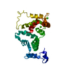


 PDBj
PDBj


 Assembly


 Mass: 507.181 Da / Num. of mol.: 1 / Source method: obtained synthetically / Formula: C10H16N5O13P3 / Comment: ATP, energy-carrying molecule*YM
 Mass: 427.201 Da / Num. of mol.: 1 / Source method: obtained synthetically / Formula: C10H15N5O10P2 / Comment: ADP, energy-carrying molecule*YM
 Mass: 18.015 Da / Num. of mol.: 184 / Source method: isolated from a natural source / Formula: H2O
 Sample preparation
 / Beamline: 5.0.3 / Wavelength: 1 Å
 Processing