 Movie
Movie Controller

[English] 日本語
 Yorodumi
- PDB-4j18: Crystal structure of H191L mutant of UDP-glucose pyrophosphorylas... -
+ Open data

- Basic information
| Entry | Database: PDB / ID: 4j18 | ||||||
|---|---|---|---|---|---|---|---|
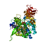
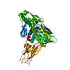
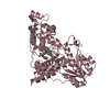
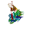
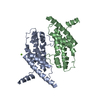
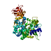
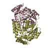
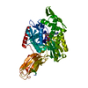
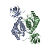
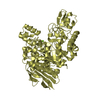
| Title | Crystal structure of H191L mutant of UDP-glucose pyrophosphorylase from Leishmania major | ||||||
 Components Components | UDP-glucose pyrophosphorylase | ||||||
 Keywords Keywords | TRANSFERASE / Rossmann-like alpha/beta/alpha sandwich fold of catalytic domain / nucleotidyltransferase / UTP / Glc-1-P | ||||||
| Function / homology |  Function and homology information Function and homology informationUTP-glucose-1-phosphate uridylyltransferase / UTP:glucose-1-phosphate uridylyltransferase activity / UDP-alpha-D-glucose metabolic process / nuclear lumen / ciliary plasm / glycogen metabolic process / cytoplasm / cytosol Similarity search - Function | ||||||
| Biological species |  Leishmania major (eukaryote) Leishmania major (eukaryote) | ||||||
| Method |  X-RAY DIFFRACTION / SYNCHROTRON / MOLECULAR REPLACEMENT / Resolution: 2.35 Å X-RAY DIFFRACTION / SYNCHROTRON / MOLECULAR REPLACEMENT / Resolution: 2.35 Å | ||||||
 Authors Authors | Fuehring, J.I. / Routier, F.H. / Lamerz, A.-C. / Baruch, P. / Gerardy-Schahn, R. / Fedorov, R. | ||||||
 Citation Citation | Journal: ACS CATALYSIS / Year: 2013 Title: Catalytic Mechanism and Allosteric Regulation of UDP-Glucose Pyrophosphorylase from Leishmania major Authors: Fuehring, J.I. / Routier, F.H. / Lamerz, A.-C. / Baruch, P. / Gerardy-Schahn, R. / Fedorov, R. | ||||||
| History |
|
- Structure visualization
| Structure viewer | Molecule: MolmilJmol/JSmol |
|---|
- Downloads & links
-Download
| PDBx/mmCIF format | 4j18.cif.gz | 113.3 KB | Display | PDBx/mmCIF format |
|---|---|---|---|---|
| PDB format | pdb4j18.ent.gz | 86.5 KB | Display | PDB format |
| PDBx/mmJSON format | 4j18.json.gz | Tree view | PDBx/mmJSON format | |
| Others |  Other downloads Other downloads |
-Validation report
| Arichive directory | https://data.pdbj.org/pub/pdb/validation_reports/j1/4j18ftp://data.pdbj.org/pub/pdb/validation_reports/j1/4j18 | HTTPS FTP |
|---|
-Related structure data
| Related structure data | 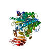 2oefS  4euj 4evk S: Starting model for refinement |
|---|---|
| Similar structure data |

-Links
 PDBj
PDBj

- Assembly
| Deposited unit | 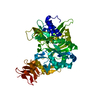
| ||||||||
|---|---|---|---|---|---|---|---|---|---|
| 1 |
| ||||||||
| Unit cell |
|
-Components
| #1: Protein | Mass: 56029.781 Da / Num. of mol.: 1 / Mutation: H191L Source method: isolated from a genetically manipulated source Source: (gene. exp.) Leishmania major (eukaryote) / Gene: UGP, LMJF_18_0990 / Production host:  References: UniProt: Q4QDU3, UTP-glucose-1-phosphate uridylyltransferase |
|---|---|
| #2: Water | ChemComp-HOH /  Mass: 18.015 Da / Num. of mol.: 309 / Source method: isolated from a natural source / Formula: H2O Mass: 18.015 Da / Num. of mol.: 309 / Source method: isolated from a natural source / Formula: H2O |
-Experimental details
-Experiment
| Experiment | Method: X-RAY DIFFRACTION / Number of used crystals: 1 |
|---|
- Sample preparation
| Crystal | Density Matthews: 3.29 Å3/Da / Density % sol: 62.58 % |
|---|---|
| Crystal grow | Temperature: 293.15 K / Method: vapor diffusion, sitting drop / pH: 5.2 Details: 10% v/v glycerol, 0.05 M citrate buffer, 1.1 M sodium citrate , pH 5.2, VAPOR DIFFUSION, SITTING DROP, temperature 293.15K |
-Data collection
| Diffraction | Mean temperature: 100 K |
|---|---|
| Diffraction source | Source: SYNCHROTRON / Site: ESRF  / Beamline: ID29 / Wavelength: 0.91985 Å / Beamline: ID29 / Wavelength: 0.91985 Å |
| Detector | Type: DECTRIS PILATUS 6M / Detector: PIXEL / Date: Nov 30, 2012 |
| Radiation | Monochromator: Liquid nitrogen cooled channel-cut silicon monochromator. Protocol: SINGLE WAVELENGTH / Monochromatic (M) / Laue (L): M / Scattering type: x-ray |
| Radiation wavelength | Wavelength: 0.91985 Å / Relative weight: 1 |
| Reflection | Resolution: 2.35→19.5 Å / Num. all: 30457 / Num. obs: 30206 / % possible obs: 99.2 % / Observed criterion σ(F): 2 / Observed criterion σ(I): 2 / Redundancy: 3.7 % / Biso Wilson estimate: 38.1 Å2 / Rsym value: 0.104 / Net I/σ(I): 9.8 |
| Reflection shell | Resolution: 2.35→2.4 Å / % possible all: 98.2 |
- Processing
| Software |
| ||||||||||||||||||||
|---|---|---|---|---|---|---|---|---|---|---|---|---|---|---|---|---|---|---|---|---|---|
| Refinement | Method to determine structure: MOLECULAR REPLACEMENT Starting model: PDB ENTRY 2OEF Resolution: 2.35→19.5 Å / Cross valid method: THROUGHOUT / σ(F): 0 / Stereochemistry target values: Engh & Huber
| ||||||||||||||||||||
| Refinement step | Cycle: LAST / Resolution: 2.35→19.5 Å
|
