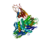
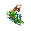
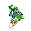
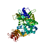
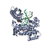
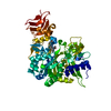
Entry Database : PDB / ID : 6eofTitle Crystal structure of AMPylated GRP78 in ADP state 78 kDa glucose-regulated protein Keywords / / / / Function / homology Function Domain/homology Component
/ / / / / / / / / / / / / / / / / / / / / / / / / / / / / / / / / / / / / / / / / / / / / / / / / / / / / Biological species Cricetulus griseus (Chinese hamster)Method / / / Resolution : 1.59 Å Authors Yan, Y. / Preissler, S. / Read, R.J. / Ron, D. Funding support Organization Grant number Country Wellcome Trust 082961/Z/07/Z Wellcome Trust 200848/Z/16/Z British Heart Foundation PG/12/41/29679
Journal : Elife / Year : 2017Title : AMPylation targets the rate-limiting step of BiP's ATPase cycle for its functional inactivation.Authors : Preissler, S. / Rohland, L. / Yan, Y. / Chen, R. / Read, R.J. / Ron, D. History Deposition Oct 9, 2017 Deposition site / Processing site Revision 1.0 Nov 1, 2017 Provider / Type Revision 1.1 Jan 17, 2024 Group / Database references / Refinement descriptionCategory chem_comp_atom / chem_comp_bond ... chem_comp_atom / chem_comp_bond / database_2 / pdbx_initial_refinement_model / software Item / _database_2.pdbx_database_accession / _software.nameRevision 1.2 Nov 20, 2024 Group / Category / pdbx_modification_feature
Show all Show less
 Movie
Movie Controller


 Open data
 Basic information
 Components
 Keywords
 Function and homology information
 Cricetulus griseus (Chinese hamster)
 X-RAY DIFFRACTION /
  Authors
 United Kingdom, 3items
  Citation
 Structure visualization
 Downloads & links
 Other downloads
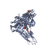


 PDBj
PDBj


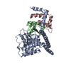
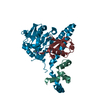
 Assembly
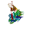


 Mass: 427.201 Da / Num. of mol.: 1 / Source method: obtained synthetically / Formula: C10H15N5O10P2 / Comment: ADP, energy-carrying molecule*YM
 Mass: 96.063 Da / Num. of mol.: 1 / Source method: obtained synthetically / Formula: SO4
 Mass: 347.221 Da / Num. of mol.: 1 / Source method: obtained synthetically / Formula: C10H14N5O7P / Comment: AMP*YM
 Mass: 18.015 Da / Num. of mol.: 172 / Source method: isolated from a natural source / Formula: H2O
 Sample preparation
 Processing