 Movie
Movie Controller

[English] 日本語
 Yorodumi
- PDB-5wzz: The SIAH E3 ubiquitin ligases promote Wnt/ beta-catenin signaling... -
+ Open data

- Basic information
| Entry | Database: PDB / ID: 5wzz | ||||||
|---|---|---|---|---|---|---|---|
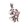
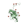
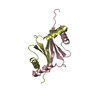
| Title | The SIAH E3 ubiquitin ligases promote Wnt/ beta-catenin signaling through mediating Wnt-induced Axin degradation | ||||||
 Components Components |
| ||||||
 Keywords Keywords | LIGASE / protein complex / WNT pathway | ||||||
| Function / homology |  Function and homology information Function and homology informationbeta-catenin destruction complex assembly / armadillo repeat domain binding / Netrin-1 signaling / head development / cell development / axial mesoderm formation / dorsal/ventral axis specification / post-anal tail morphogenesis / beta-catenin destruction complex / APC truncation mutants have impaired AXIN binding ...beta-catenin destruction complex assembly / armadillo repeat domain binding / Netrin-1 signaling / head development / cell development / axial mesoderm formation / dorsal/ventral axis specification / post-anal tail morphogenesis / beta-catenin destruction complex / APC truncation mutants have impaired AXIN binding / AXIN missense mutants destabilize the destruction complex / Truncations of AMER1 destabilize the destruction complex / Beta-catenin phosphorylation cascade / Signaling by GSK3beta mutants / CTNNB1 S33 mutants aren't phosphorylated / CTNNB1 S37 mutants aren't phosphorylated / CTNNB1 S45 mutants aren't phosphorylated / CTNNB1 T41 mutants aren't phosphorylated / I-SMAD binding / Wnt signalosome / negative regulation of protein metabolic process / Disassembly of the destruction complex and recruitment of AXIN to the membrane / ubiquitin conjugating enzyme binding / epigenetic programming in the zygotic pronuclei / positive regulation of ubiquitin-dependent protein catabolic process / nucleocytoplasmic transport / RUNX1 regulates transcription of genes involved in WNT signaling / RUNX1 regulates estrogen receptor mediated transcription / negative regulation of fat cell differentiation / positive regulation of transforming growth factor beta receptor signaling pathway / SMAD binding / R-SMAD binding / negative regulation of transcription elongation by RNA polymerase II / lateral plasma membrane / anatomical structure morphogenesis / canonical Wnt signaling pathway / ubiquitin-like ligase-substrate adaptor activity / positive regulation of intrinsic apoptotic signaling pathway / cytoplasmic microtubule organization / signaling adaptor activity / axon guidance / positive regulation of protein ubiquitination / protein catabolic process / protein serine/threonine kinase activator activity / protein serine/threonine kinase binding / cell periphery / TCF dependent signaling in response to WNT / Degradation of AXIN / sensory perception of sound / negative regulation of canonical Wnt signaling pathway / RING-type E3 ubiquitin transferase / beta-catenin binding / positive regulation of JNK cascade / protein destabilization / Degradation of beta-catenin by the destruction complex / protein polyubiquitination / p53 binding / ubiquitin-protein transferase activity / positive regulation of protein catabolic process / ubiquitin protein ligase activity / nervous system development / positive regulation of proteasomal ubiquitin-dependent protein catabolic process / Antigen processing: Ubiquitination & Proteasome degradation / microtubule cytoskeleton / protein-containing complex assembly / neuron apoptotic process / cytoplasmic vesicle / spermatogenesis / cell cortex / Estrogen-dependent gene expression / amyloid fibril formation / molecular adaptor activity / ubiquitin-dependent protein catabolic process / in utero embryonic development / proteasome-mediated ubiquitin-dependent protein catabolic process / early endosome / Ub-specific processing proteases / protein ubiquitination / positive regulation of apoptotic process / Amyloid fiber formation / negative regulation of gene expression / apoptotic process / ubiquitin protein ligase binding / protein kinase binding / nucleolus / perinuclear region of cytoplasm / enzyme binding / protein homodimerization activity / zinc ion binding / identical protein binding / nucleus / plasma membrane / cytoplasm / cytosol Similarity search - Function | ||||||
| Biological species |  Homo sapiens (human) Homo sapiens (human) | ||||||
| Method |  X-RAY DIFFRACTION / SYNCHROTRON / MOLECULAR REPLACEMENT / Resolution: 2.103 Å X-RAY DIFFRACTION / SYNCHROTRON / MOLECULAR REPLACEMENT / Resolution: 2.103 Å | ||||||
 Authors Authors | Ji, L. / Jiang, B. / Jiang, X. / Charlat, O. / Chen, A. / Mickanin, C. / Bauer, A. / Xu, W. / Yan, X.-X. / Cong, F. | ||||||
 Citation Citation | Journal: Genes Dev. / Year: 2017 Title: The SIAH E3 ubiquitin ligases promote Wnt/ beta-catenin signaling through mediating Wnt-induced Axin degradation Authors: Ji, L. / Jiang, B. / Jiang, X. / Charlat, O. / Chen, A. / Mickanin, C. / Bauer, A. / Xu, W. / Yan, X.-X. / Cong, F. | ||||||
| History |
|
- Structure visualization
| Structure viewer | Molecule: MolmilJmol/JSmol |
|---|
- Downloads & links
-Download
| PDBx/mmCIF format | 5wzz.cif.gz | 333.5 KB | Display | PDBx/mmCIF format |
|---|---|---|---|---|
| PDB format | pdb5wzz.ent.gz | 272.7 KB | Display | PDB format |
| PDBx/mmJSON format | 5wzz.json.gz | Tree view | PDBx/mmJSON format | |
| Others |  Other downloads Other downloads |
-Validation report
| Arichive directory | https://data.pdbj.org/pub/pdb/validation_reports/wz/5wzzftp://data.pdbj.org/pub/pdb/validation_reports/wz/5wzz | HTTPS FTP |
|---|
-Related structure data
| Related structure data | 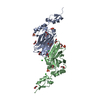 4ca1S S: Starting model for refinement |
|---|---|
| Similar structure data |


-Links
 PDBj
PDBj


- Assembly
| Deposited unit | 
| ||||||||
|---|---|---|---|---|---|---|---|---|---|
| 1 | 
| ||||||||
| 2 | 
| ||||||||
| 3 | 
| ||||||||
| 4 | 
| ||||||||
| Unit cell |
|
-Components
| #1: Protein | Mass: 21409.508 Da / Num. of mol.: 4 / Fragment: UNP residues 93-282 Source method: isolated from a genetically manipulated source Source: (gene. exp.) Homo sapiens (human) / Gene: SIAH1, HUMSIAH / Production host:  References: UniProt: Q8IUQ4, Ligases; Forming carbon-nitrogen bonds; Acid-amino-acid ligases (peptide synthases) #2: Protein/peptide | Mass: 2472.856 Da / Num. of mol.: 4 / Fragment: UNP residues 375-394 Source method: isolated from a genetically manipulated source Source: (gene. exp.) Homo sapiens (human) / Gene: AXIN1, AXIN / Production host: #3: Chemical | ChemComp-ZN /   Mass: 65.409 Da / Num. of mol.: 8 / Source method: obtained synthetically / Formula: Zn Mass: 65.409 Da / Num. of mol.: 8 / Source method: obtained synthetically / Formula: Zn#4: Water | ChemComp-HOH / |  Mass: 18.015 Da / Num. of mol.: 216 / Source method: isolated from a natural source / Formula: H2O Mass: 18.015 Da / Num. of mol.: 216 / Source method: isolated from a natural source / Formula: H2O |
|---|
-Experimental details
-Experiment
| Experiment | Method: X-RAY DIFFRACTION / Number of used crystals: 1 |
|---|
- Sample preparation
| Crystal | Density Matthews: 2.18 Å3/Da / Density % sol: 43.62 % |
|---|---|
| Crystal grow | Temperature: 277 K / Method: vapor diffusion, hanging drop / pH: 8 / Details: 25% (w/v) PEG3350, 200 mM MgCl2, 100 mM Tris-Hcl |
-Data collection
| Diffraction | Mean temperature: 100 K |
|---|---|
| Diffraction source | Source: SYNCHROTRON / Site: SSRF  / Beamline: BL19U1 / Wavelength: 1 Å / Beamline: BL19U1 / Wavelength: 1 Å |
| Detector | Type: DECTRIS PILATUS3 R CdTe 300K / Detector: PIXEL / Date: Jun 28, 2016 |
| Radiation | Protocol: SINGLE WAVELENGTH / Monochromatic (M) / Laue (L): M / Scattering type: x-ray |
| Radiation wavelength | Wavelength: 1 Å / Relative weight: 1 |
| Reflection | Resolution: 2.1→40 Å / Num. obs: 92372 / % possible obs: 99.6 % / Redundancy: 6.3 % / Net I/σ(I): 30 |
- Processing
| Software |
| |||||||||||||||||||||||||||||||||||||||||||||||||||||||||||||||||||||||||||||||||||||||||||||||||||||||||||||||||||||||
|---|---|---|---|---|---|---|---|---|---|---|---|---|---|---|---|---|---|---|---|---|---|---|---|---|---|---|---|---|---|---|---|---|---|---|---|---|---|---|---|---|---|---|---|---|---|---|---|---|---|---|---|---|---|---|---|---|---|---|---|---|---|---|---|---|---|---|---|---|---|---|---|---|---|---|---|---|---|---|---|---|---|---|---|---|---|---|---|---|---|---|---|---|---|---|---|---|---|---|---|---|---|---|---|---|---|---|---|---|---|---|---|---|---|---|---|---|---|---|---|---|
| Refinement | Method to determine structure: MOLECULAR REPLACEMENT Starting model: 4ca1 Resolution: 2.103→38.728 Å / SU ML: 0.22 / Cross valid method: FREE R-VALUE / σ(F): 1.35 / Phase error: 22.28 / Stereochemistry target values: ML
| |||||||||||||||||||||||||||||||||||||||||||||||||||||||||||||||||||||||||||||||||||||||||||||||||||||||||||||||||||||||
| Solvent computation | Shrinkage radii: 0.9 Å / VDW probe radii: 1.11 Å / Solvent model: FLAT BULK SOLVENT MODEL | |||||||||||||||||||||||||||||||||||||||||||||||||||||||||||||||||||||||||||||||||||||||||||||||||||||||||||||||||||||||
| Refinement step | Cycle: LAST / Resolution: 2.103→38.728 Å
| |||||||||||||||||||||||||||||||||||||||||||||||||||||||||||||||||||||||||||||||||||||||||||||||||||||||||||||||||||||||
| Refine LS restraints |
| |||||||||||||||||||||||||||||||||||||||||||||||||||||||||||||||||||||||||||||||||||||||||||||||||||||||||||||||||||||||
| LS refinement shell |
|
