 Movie
Movie Controller

[English] 日本語
 Yorodumi
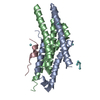
- PDB-4mko: Crystal structure of the monomeric, cleaved form of the Pore-Form... -
+ Open data

- Basic information
| Entry | Database: PDB / ID: 4mko | ||||||
|---|---|---|---|---|---|---|---|
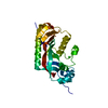
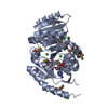
| Title | Crystal structure of the monomeric, cleaved form of the Pore-Forming Toxin Monalysin | ||||||
 Components Components | Monalysin | ||||||
 Keywords Keywords | TOXIN / Pore-Forming Toxin | ||||||
| Function / homology |  Function and homology information Function and homology informationhemolysis in another organism / porin activity / pore complex / monoatomic ion transport / protein homooligomerization / toxin activity / host cell plasma membrane / extracellular region Similarity search - Function | ||||||
| Biological species |  Pseudomonas entomophila (bacteria) Pseudomonas entomophila (bacteria) | ||||||
| Method |  X-RAY DIFFRACTION / SYNCHROTRON / MOLECULAR REPLACEMENT / Resolution: 1.7 Å X-RAY DIFFRACTION / SYNCHROTRON / MOLECULAR REPLACEMENT / Resolution: 1.7 Å | ||||||
 Authors Authors | Leone, P. / Roussel, A. | ||||||
 Citation Citation | Journal: J Biol Chem / Year: 2015 Title: X-ray and Cryo-electron Microscopy Structures of Monalysin Pore-forming Toxin Reveal Multimerization of the Pro-form. Authors: Philippe Leone / Cecilia Bebeacua / Onya Opota / Christine Kellenberger / Bruno Klaholz / Igor Orlov / Christian Cambillau / Bruno Lemaitre / Alain Roussel /   Abstract: β-Barrel pore-forming toxins (β-PFT), a large family of bacterial toxins, are generally secreted as water-soluble monomers and can form oligomeric pores in membranes following proteolytic cleavage ...β-Barrel pore-forming toxins (β-PFT), a large family of bacterial toxins, are generally secreted as water-soluble monomers and can form oligomeric pores in membranes following proteolytic cleavage and interaction with cell surface receptors. Monalysin has been recently identified as a β-PFT that contributes to the virulence of Pseudomonas entomophila against Drosophila. It is secreted as a pro-protein that becomes active upon cleavage. Here we report the crystal and cryo-electron microscopy structure of the pro-form of Monalysin as well as the crystal structures of the cleaved form and of an inactive mutant lacking the membrane-spanning region. The overall structure of Monalysin displays an elongated shape, which resembles those of β-pore-forming toxins, such as Aerolysin, but is devoid of a receptor-binding domain. X-ray crystallography, cryo-electron microscopy, and light-scattering studies show that pro-Monalysin forms a stable doughnut-like 18-mer complex composed of two disk-shaped nonamers held together by N-terminal swapping of the pro-peptides. This observation is in contrast with the monomeric pro-form of the other β-PFTs that are receptor-dependent for membrane interaction. The membrane-spanning region of pro-Monalysin is fully buried in the center of the doughnut, suggesting that upon cleavage of pro-peptides, the two disk-shaped nonamers can, and have to, dissociate to leave the transmembrane segments free to deploy and lead to pore formation. In contrast with other toxins, the delivery of 18 subunits at once, nearby the cell surface, may be used to bypass the requirement of receptor-dependent concentration to reach the threshold for oligomerization into the pore-forming complex. | ||||||
| History |
|
- Structure visualization
| Structure viewer | Molecule: MolmilJmol/JSmol |
|---|
- Downloads & links
-Download
| PDBx/mmCIF format | 4mko.cif.gz | 233.6 KB | Display | PDBx/mmCIF format |
|---|---|---|---|---|
| PDB format | pdb4mko.ent.gz | 186 KB | Display | PDB format |
| PDBx/mmJSON format | 4mko.json.gz | Tree view | PDBx/mmJSON format | |
| Others |  Other downloads Other downloads |
-Validation report
| Arichive directory | https://data.pdbj.org/pub/pdb/validation_reports/mk/4mkoftp://data.pdbj.org/pub/pdb/validation_reports/mk/4mko | HTTPS FTP |
|---|
-Related structure data
| Related structure data | 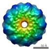 2698C 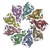 4mjtSC  4mkqC S: Starting model for refinement C: citing same article ( |
|---|---|
| Similar structure data |

-Links
 PDBj
PDBj


- Assembly
| Deposited unit | 
| ||||||||
|---|---|---|---|---|---|---|---|---|---|
| 1 | 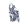
| ||||||||
| 2 | 
| ||||||||
| 3 | 
| ||||||||
| 4 | 
| ||||||||
| Unit cell |
|
-Components
| #1: Protein | Mass: 26458.146 Da / Num. of mol.: 4 / Fragment: UNP residues 36-271 Source method: isolated from a genetically manipulated source Source: (gene. exp.) Pseudomonas entomophila (bacteria) / Strain: L48 / Gene: PSEEN3174 / Production host: #2: Chemical | ChemComp-HG /   Mass: 200.590 Da / Num. of mol.: 8 / Source method: obtained synthetically / Formula: Hg Mass: 200.590 Da / Num. of mol.: 8 / Source method: obtained synthetically / Formula: Hg#3: Chemical | ChemComp-ZN /   Mass: 65.409 Da / Num. of mol.: 8 / Source method: obtained synthetically / Formula: Zn Mass: 65.409 Da / Num. of mol.: 8 / Source method: obtained synthetically / Formula: Zn#4: Chemical | ChemComp-ACT /   Mass: 59.044 Da / Num. of mol.: 13 / Source method: obtained synthetically / Formula: C2H3O2 Mass: 59.044 Da / Num. of mol.: 13 / Source method: obtained synthetically / Formula: C2H3O2#5: Water | ChemComp-HOH / |  Mass: 18.015 Da / Num. of mol.: 1318 / Source method: isolated from a natural source / Formula: H2O Mass: 18.015 Da / Num. of mol.: 1318 / Source method: isolated from a natural source / Formula: H2O |
|---|
-Experimental details
-Experiment
| Experiment | Method: X-RAY DIFFRACTION / Number of used crystals: 1 |
|---|
- Sample preparation
| Crystal | Density Matthews: 2.71 Å3/Da / Density % sol: 54.67 % |
|---|---|
| Crystal grow | Temperature: 293 K / Method: vapor diffusion, hanging drop / pH: 4.6 Details: 0.1M sodium acetate, pH 4.6, 0.13M zinc acetate, 0.6-1.1M ammonium acetate, 2-7% PEG800, 1.5mM HgCl2, VAPOR DIFFUSION, HANGING DROP, temperature 293K |
-Data collection
| Diffraction | Mean temperature: 100 K |
|---|---|
| Diffraction source | Source: SYNCHROTRON / Site: ESRF / Beamline: ID14-4 / Wavelength: 0.9393 Å |
| Radiation | Protocol: SINGLE WAVELENGTH / Monochromatic (M) / Laue (L): M / Scattering type: x-ray |
| Radiation wavelength | Wavelength: 0.9393 Å / Relative weight: 1 |
| Reflection | Resolution: 1.7→30 Å / Num. all: 126982 / Num. obs: 126982 / % possible obs: 100 % / Observed criterion σ(F): 0 / Observed criterion σ(I): 0 / Redundancy: 6.7 % / Biso Wilson estimate: 14.11 Å2 / Rmerge(I) obs: 0.093 / Net I/σ(I): 12.9 |
| Reflection shell | Resolution: 1.7→1.79 Å / Redundancy: 6.8 % / Rmerge(I) obs: 0.445 / Mean I/σ(I) obs: 3.9 / Num. unique all: 18400 / % possible all: 100 |
- Processing
| Software | Name: BUSTER / Version: 2.11.4 / Classification: refinement | ||||||||||||||||||||||||||||||||||||||||||||||||||||||||||||||||||||||||||||||
|---|---|---|---|---|---|---|---|---|---|---|---|---|---|---|---|---|---|---|---|---|---|---|---|---|---|---|---|---|---|---|---|---|---|---|---|---|---|---|---|---|---|---|---|---|---|---|---|---|---|---|---|---|---|---|---|---|---|---|---|---|---|---|---|---|---|---|---|---|---|---|---|---|---|---|---|---|---|---|---|
| Refinement | Method to determine structure: MOLECULAR REPLACEMENT Starting model: PBD entry 4MJT Resolution: 1.7→29.78 Å / Cor.coef. Fo:Fc: 0.9404 / Cor.coef. Fo:Fc free: 0.9283 / SU R Cruickshank DPI: 0.096 / Cross valid method: THROUGHOUT / σ(F): 0
| ||||||||||||||||||||||||||||||||||||||||||||||||||||||||||||||||||||||||||||||
| Displacement parameters | Biso mean: 17.56 Å2
| ||||||||||||||||||||||||||||||||||||||||||||||||||||||||||||||||||||||||||||||
| Refine analyze | Luzzati coordinate error obs: 0.185 Å | ||||||||||||||||||||||||||||||||||||||||||||||||||||||||||||||||||||||||||||||
| Refinement step | Cycle: LAST / Resolution: 1.7→29.78 Å
| ||||||||||||||||||||||||||||||||||||||||||||||||||||||||||||||||||||||||||||||
| Refine LS restraints |
| ||||||||||||||||||||||||||||||||||||||||||||||||||||||||||||||||||||||||||||||
| LS refinement shell | Resolution: 1.7→1.74 Å / Total num. of bins used: 20
|
