- PDB-4mjt: Crystal structure of the oligomeric pore-forming toxin pro-Monalysin -
+
Open data
ID or keywords:
Loading...
-
Basic information
Entry
Database: PDB / ID: 4mjt
Title
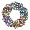
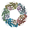
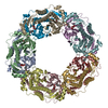
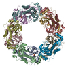
Crystal structure of the oligomeric pore-forming toxin pro-Monalysin
Components
MONALYSIN
Monalysin
Keywords
TOXIN / Pore-Forming Toxin
Function / homology
Function and homology information
hemolysis in another organism / porin activity / pore complex / monoatomic ion transport / protein homooligomerization / toxin activity / host cell plasma membrane / extracellular region Similarity search - Function
Journal: J Biol Chem / Year: 2015 Title: X-ray and Cryo-electron Microscopy Structures of Monalysin Pore-forming Toxin Reveal Multimerization of the Pro-form. Authors: Philippe Leone / Cecilia Bebeacua / Onya Opota / Christine Kellenberger / Bruno Klaholz / Igor Orlov / Christian Cambillau / Bruno Lemaitre / Alain Roussel / Abstract: β-Barrel pore-forming toxins (β-PFT), a large family of bacterial toxins, are generally secreted as water-soluble monomers and can form oligomeric pores in membranes following proteolytic cleavage ...β-Barrel pore-forming toxins (β-PFT), a large family of bacterial toxins, are generally secreted as water-soluble monomers and can form oligomeric pores in membranes following proteolytic cleavage and interaction with cell surface receptors. Monalysin has been recently identified as a β-PFT that contributes to the virulence of Pseudomonas entomophila against Drosophila. It is secreted as a pro-protein that becomes active upon cleavage. Here we report the crystal and cryo-electron microscopy structure of the pro-form of Monalysin as well as the crystal structures of the cleaved form and of an inactive mutant lacking the membrane-spanning region. The overall structure of Monalysin displays an elongated shape, which resembles those of β-pore-forming toxins, such as Aerolysin, but is devoid of a receptor-binding domain. X-ray crystallography, cryo-electron microscopy, and light-scattering studies show that pro-Monalysin forms a stable doughnut-like 18-mer complex composed of two disk-shaped nonamers held together by N-terminal swapping of the pro-peptides. This observation is in contrast with the monomeric pro-form of the other β-PFTs that are receptor-dependent for membrane interaction. The membrane-spanning region of pro-Monalysin is fully buried in the center of the doughnut, suggesting that upon cleavage of pro-peptides, the two disk-shaped nonamers can, and have to, dissociate to leave the transmembrane segments free to deploy and lead to pore formation. In contrast with other toxins, the delivery of 18 subunits at once, nearby the cell surface, may be used to bypass the requirement of receptor-dependent concentration to reach the threshold for oligomerization into the pore-forming complex.
Mass: 18.015 Da / Num. of mol.: 656 / Source method: isolated from a natural source / Formula: H2O
Compound details
AUTHORS STATE THAT PEPTIDE CHAIN (RESIDUES 9-35) AND PROTEIN CHAIN (RESIDUES 36-271) BELONG TO A ...AUTHORS STATE THAT PEPTIDE CHAIN (RESIDUES 9-35) AND PROTEIN CHAIN (RESIDUES 36-271) BELONG TO A SAME SINGLE CHAIN WITHOUT BEEN CLEAVED BETWEEN RESIDUES 35 AND 36. AUTHORS HAVE STRONG EVIDENCE OF THE DOMAIN SWAPPING BUT NO EXPERIMENTAL DATA COULD PERMIT TO ASSIGN THE CHAINS UNAMBIGUOUSLY. THEREFORE, THEY WERE ASSIGNED DISTINCT CHAIN IDS.
-
Experimental details
-
Experiment
Experiment
Method: X-RAY DIFFRACTION / Number of used crystals: 1
-
Sample preparation
Crystal
Density Matthews: 2.73 Å3/Da / Density % sol: 54.89 %
In the structure databanks used in Yorodumi, some data are registered as the other names, "COVID-19 virus" and "2019-nCoV". Here are the details of the virus and the list of structure data.
Jan 31, 2019. EMDB accession codes are about to change! (news from PDBe EMDB page)
EMDB accession codes are about to change! (news from PDBe EMDB page)
The allocation of 4 digits for EMDB accession codes will soon come to an end. Whilst these codes will remain in use, new EMDB accession codes will include an additional digit and will expand incrementally as the available range of codes is exhausted. The current 4-digit format prefixed with “EMD-” (i.e. EMD-XXXX) will advance to a 5-digit format (i.e. EMD-XXXXX), and so on. It is currently estimated that the 4-digit codes will be depleted around Spring 2019, at which point the 5-digit format will come into force.
The EM Navigator/Yorodumi systems omit the EMD- prefix.
Related info.:Q: What is EMD? / ID/Accession-code notation in Yorodumi/EM Navigator
Yorodumi is a browser for structure data from EMDB, PDB, SASBDB, etc.
This page is also the successor to EM Navigator detail page, and also detail information page/front-end page for Omokage search.
The word "yorodu" (or yorozu) is an old Japanese word meaning "ten thousand". "mi" (miru) is to see.
Related info.:EMDB / PDB / SASBDB / Comparison of 3 databanks / Yorodumi Search / Aug 31, 2016. New EM Navigator & Yorodumi / Yorodumi Papers / Jmol/JSmol / Function and homology information / Changes in new EM Navigator and Yorodumi
 Movie
Movie Controller

 Yorodumi
 Open data
 Basic information
 Components
 Keywords
 Function and homology information
 Pseudomonas entomophila (bacteria)
 X-RAY DIFFRACTION /
  Authors
 Citation

 Structure visualization
 Downloads & links
 Other downloads
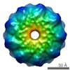


 PDBj
PDBj Assembly

 Mass: 65.409 Da / Num. of mol.: 27 / Source method: obtained synthetically / Formula: Zn
 Mass: 18.015 Da / Num. of mol.: 656 / Source method: isolated from a natural source / Formula: H2O
 Sample preparation
 Processing