 Movie
Movie Controller

[English] 日本語
 Yorodumi
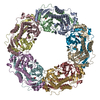
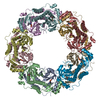
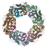
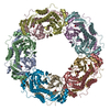
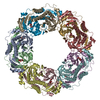
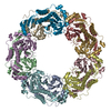
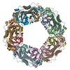
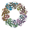
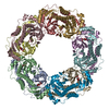
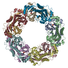
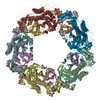
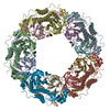
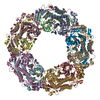
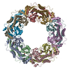
- PDB-7c89: Peroxiredoxin from Aeropyrum pernix K1 (ApPrx) C50S/F80C/C207S/C2... -
+ Open data

- Basic information
| Entry | Database: PDB / ID: 7c89 | |||||||||
|---|---|---|---|---|---|---|---|---|---|---|
| Title | Peroxiredoxin from Aeropyrum pernix K1 (ApPrx) C50S/F80C/C207S/C213S mutant modified with 2-bromoacetophenone(Ph@ApPrx*) | |||||||||
 Components Components | Peroxiredoxin | |||||||||
 Keywords Keywords | OXIDOREDUCTASE / Peroxiredoxin | |||||||||
| Function / homology |  Function and homology information Function and homology informationperoxiredoxin activity / thioredoxin-dependent peroxiredoxin / thioredoxin peroxidase activity / antioxidant activity / cell redox homeostasis / hydrogen peroxide catabolic process / cellular response to hydrogen peroxide / identical protein binding / cytosol Similarity search - Function | |||||||||
| Biological species |   Aeropyrum pernix K1 (archaea) Aeropyrum pernix K1 (archaea) | |||||||||
| Method |  X-RAY DIFFRACTION / SYNCHROTRON / MOLECULAR REPLACEMENT / Resolution: 2.1 Å X-RAY DIFFRACTION / SYNCHROTRON / MOLECULAR REPLACEMENT / Resolution: 2.1 Å | |||||||||
 Authors Authors | Himiyama, T. / Nakamura, T. | |||||||||
| Funding support |  Japan, 2items Japan, 2items
| |||||||||
 Citation Citation | Journal: Bioconjug.Chem. / Year: 2021 Title: Rebuilding Ring-Type Assembly of Peroxiredoxin by Chemical Modification. Authors: Himiyama, T. / Tsuchiya, Y. / Yonezawa, Y. / Nakamura, T. | |||||||||
| History |
|
- Structure visualization
| Structure viewer | Molecule: MolmilJmol/JSmol |
|---|
- Downloads & links
-Download
| PDBx/mmCIF format | 7c89.cif.gz | 525.4 KB | Display | PDBx/mmCIF format |
|---|---|---|---|---|
| PDB format | pdb7c89.ent.gz | 419.7 KB | Display | PDB format |
| PDBx/mmJSON format | 7c89.json.gz | Tree view | PDBx/mmJSON format | |
| Others |  Other downloads Other downloads |
-Validation report
| Arichive directory | https://data.pdbj.org/pub/pdb/validation_reports/c8/7c89ftp://data.pdbj.org/pub/pdb/validation_reports/c8/7c89 | HTTPS FTP |
|---|
-Related structure data
| Related structure data |  7c87C 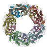 7c8aC  7cqjC  6krkS S: Starting model for refinement C: citing same article ( |
|---|---|
| Similar structure data |


-Links
 PDBj
PDBj


- Assembly
| Deposited unit | 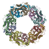
| ||||||||
|---|---|---|---|---|---|---|---|---|---|
| 1 |
| ||||||||
| Unit cell |
|
-Components
| #1: Protein | Mass: 28651.797 Da / Num. of mol.: 10 / Mutation: C50S, F80C, C207S, C213S Source method: isolated from a genetically manipulated source Source: (gene. exp.) Aeropyrum pernix K1 (archaea) / Strain: K1 / Gene: APE_2278 / Production host:  #2: Chemical | ChemComp-FLC /   Mass: 189.100 Da / Num. of mol.: 10 / Source method: obtained synthetically / Formula: C6H5O7 Mass: 189.100 Da / Num. of mol.: 10 / Source method: obtained synthetically / Formula: C6H5O7#3: Chemical | ChemComp-FL0 / 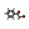  Mass: 199.045 Da / Num. of mol.: 10 / Source method: obtained synthetically / Formula: C8H7BrO / Feature type: SUBJECT OF INVESTIGATION Mass: 199.045 Da / Num. of mol.: 10 / Source method: obtained synthetically / Formula: C8H7BrO / Feature type: SUBJECT OF INVESTIGATION#4: Water | ChemComp-HOH / |  Mass: 18.015 Da / Num. of mol.: 1146 / Source method: isolated from a natural source / Formula: H2O Mass: 18.015 Da / Num. of mol.: 1146 / Source method: isolated from a natural source / Formula: H2OHas ligand of interest | Y | Has protein modification | Y | |
|---|
-Experimental details
-Experiment
| Experiment | Method: X-RAY DIFFRACTION / Number of used crystals: 1 |
|---|
- Sample preparation
| Crystal | Density Matthews: 2.65 Å3/Da / Density % sol: 53.61 % |
|---|---|
| Crystal grow | Temperature: 293 K / Method: vapor diffusion, hanging drop Details: The reservoir solution contained 0.10 M sodium citrate (pH5.5) 0.20 M lithium sulfate 15% (v/v) reagent alcohol. |
-Data collection
| Diffraction | Mean temperature: 100 K / Serial crystal experiment: N |
|---|---|
| Diffraction source | Source: SYNCHROTRON / Site: SPring-8 / Beamline: BL45XU / Wavelength: 1 Å |
| Detector | Type: DECTRIS PILATUS 6M / Detector: PIXEL / Date: Oct 12, 2019 |
| Radiation | Protocol: SINGLE WAVELENGTH / Monochromatic (M) / Laue (L): M / Scattering type: x-ray |
| Radiation wavelength | Wavelength: 1 Å / Relative weight: 1 |
| Reflection | Resolution: 2.1→50 Å / Num. obs: 168919 / % possible obs: 98.3 % / Redundancy: 3.6 % / CC1/2: 0.996 / Rmerge(I) obs: 0.08 / Net I/σ(I): 10.1 |
| Reflection shell | Resolution: 2.1→2.14 Å / Rmerge(I) obs: 0.391 / Mean I/σ(I) obs: 3 / Num. unique obs: 8244 / CC1/2: 0.889 |
- Processing
| Software |
| |||||||||||||||||||||||||||||||||||||||||||||||||||||||||||||||||||||||||||||||||||||||||||||||||||||||||||||||||||||||||||||||||||||||||||||||||||||||||||
|---|---|---|---|---|---|---|---|---|---|---|---|---|---|---|---|---|---|---|---|---|---|---|---|---|---|---|---|---|---|---|---|---|---|---|---|---|---|---|---|---|---|---|---|---|---|---|---|---|---|---|---|---|---|---|---|---|---|---|---|---|---|---|---|---|---|---|---|---|---|---|---|---|---|---|---|---|---|---|---|---|---|---|---|---|---|---|---|---|---|---|---|---|---|---|---|---|---|---|---|---|---|---|---|---|---|---|---|---|---|---|---|---|---|---|---|---|---|---|---|---|---|---|---|---|---|---|---|---|---|---|---|---|---|---|---|---|---|---|---|---|---|---|---|---|---|---|---|---|---|---|---|---|---|---|---|---|
| Refinement | Method to determine structure: MOLECULAR REPLACEMENT Starting model: 6KRK Resolution: 2.1→49.168 Å / Cor.coef. Fo:Fc: 0.964 / Cor.coef. Fo:Fc free: 0.939 / SU B: 4.406 / SU ML: 0.115 / Cross valid method: THROUGHOUT / ESU R: 0.179 / ESU R Free: 0.163 Details: Hydrogens have been added in their riding positions
| |||||||||||||||||||||||||||||||||||||||||||||||||||||||||||||||||||||||||||||||||||||||||||||||||||||||||||||||||||||||||||||||||||||||||||||||||||||||||||
| Solvent computation | Ion probe radii: 0.8 Å / Shrinkage radii: 0.8 Å / VDW probe radii: 1.2 Å / Solvent model: BABINET MODEL PLUS MASK | |||||||||||||||||||||||||||||||||||||||||||||||||||||||||||||||||||||||||||||||||||||||||||||||||||||||||||||||||||||||||||||||||||||||||||||||||||||||||||
| Displacement parameters | Biso mean: 33.297 Å2
| |||||||||||||||||||||||||||||||||||||||||||||||||||||||||||||||||||||||||||||||||||||||||||||||||||||||||||||||||||||||||||||||||||||||||||||||||||||||||||
| Refinement step | Cycle: LAST / Resolution: 2.1→49.168 Å
| |||||||||||||||||||||||||||||||||||||||||||||||||||||||||||||||||||||||||||||||||||||||||||||||||||||||||||||||||||||||||||||||||||||||||||||||||||||||||||
| Refine LS restraints |
| |||||||||||||||||||||||||||||||||||||||||||||||||||||||||||||||||||||||||||||||||||||||||||||||||||||||||||||||||||||||||||||||||||||||||||||||||||||||||||
| LS refinement shell |
|
