 Movie
Movie Controller

[English] 日本語
 Yorodumi
- PDB-2e2g: Crystal structure of archaeal peroxiredoxin, thioredoxin peroxida... -
+ Open data

- Basic information
| Entry | Database: PDB / ID: 2e2g | ||||||
|---|---|---|---|---|---|---|---|
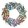
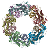
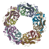
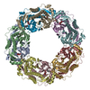
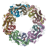
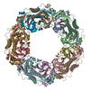
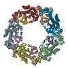
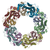
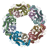
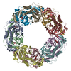
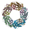
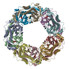
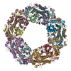
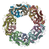
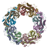
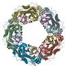
| Title | Crystal structure of archaeal peroxiredoxin, thioredoxin peroxidase from Aeropyrum pernix K1 (pre-oxidation form) | ||||||
 Components Components | Probable peroxiredoxin | ||||||
 Keywords Keywords | OXIDOREDUCTASE / thioredoxin peroxidase / cysteine sulfenic acid / cysteine sulfinic acid / cysteine sulfonic acid / hypervalent sulfur compound / peroxidatic cysteine | ||||||
| Function / homology |  Function and homology information Function and homology informationperoxiredoxin activity / thioredoxin-dependent peroxiredoxin / thioredoxin peroxidase activity / antioxidant activity / cell redox homeostasis / hydrogen peroxide catabolic process / cellular response to hydrogen peroxide / identical protein binding / cytosol Similarity search - Function | ||||||
| Biological species |   Aeropyrum pernix (archaea) Aeropyrum pernix (archaea) | ||||||
| Method |  X-RAY DIFFRACTION / SYNCHROTRON / MOLECULAR REPLACEMENT / Resolution: 2.4 Å X-RAY DIFFRACTION / SYNCHROTRON / MOLECULAR REPLACEMENT / Resolution: 2.4 Å | ||||||
 Authors Authors | Nakamura, T. / Yamamoto, T. / Abe, M. / Matsumura, H. / Hagihara, Y. / Goto, T. / Yamaguchi, T. / Inoue, T. | ||||||
 Citation Citation | Journal: Proc.Natl.Acad.Sci.Usa / Year: 2008 Title: Oxidation of archaeal peroxiredoxin involves a hypervalent sulfur intermediate Authors: Nakamura, T. / Yamamoto, T. / Abe, M. / Matsumura, H. / Hagihara, Y. / Goto, T. / Yamaguchi, T. / Inoue, T. | ||||||
| History |
|
- Structure visualization
| Structure viewer | Molecule: MolmilJmol/JSmol |
|---|
- Downloads & links
-Download
| PDBx/mmCIF format | 2e2g.cif.gz | 476.6 KB | Display | PDBx/mmCIF format |
|---|---|---|---|---|
| PDB format | pdb2e2g.ent.gz | 395.1 KB | Display | PDB format |
| PDBx/mmJSON format | 2e2g.json.gz | Tree view | PDBx/mmJSON format | |
| Others |  Other downloads Other downloads |
-Validation report
| Arichive directory | https://data.pdbj.org/pub/pdb/validation_reports/e2/2e2gftp://data.pdbj.org/pub/pdb/validation_reports/e2/2e2g | HTTPS FTP |
|---|
-Related structure data
| Related structure data |  2e2mC  2nvlC  2zctC  1x0rS C: citing same article ( S: Starting model for refinement |
|---|---|
| Similar structure data |
-Links
 PDBj
PDBj- Assembly
| Deposited unit | 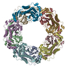
| ||||||||
|---|---|---|---|---|---|---|---|---|---|
| 1 |
| ||||||||
| Unit cell |
|
-Components
| #1: Protein | Mass: 28727.957 Da / Num. of mol.: 10 / Mutation: C207S Source method: isolated from a genetically manipulated source Source: (gene. exp.) Aeropyrum pernix (archaea) / Strain: K1 / Plasmid: pET3d / Production host:  #2: Water | ChemComp-HOH / |  Mass: 18.015 Da / Num. of mol.: 542 / Source method: isolated from a natural source / Formula: H2O Mass: 18.015 Da / Num. of mol.: 542 / Source method: isolated from a natural source / Formula: H2O |
|---|
-Experimental details
-Experiment
| Experiment | Method: X-RAY DIFFRACTION / Number of used crystals: 1 |
|---|
- Sample preparation
| Crystal | Density Matthews: 2.64 Å3/Da / Density % sol: 53.42 % |
|---|---|
| Crystal grow | Temperature: 293 K / Method: vapor diffusion, hanging drop / pH: 4.8 Details: 0.1M Acetate buffer pH4.8, 0.2M calcium chloride, 10%(v/v)isopropanol, VAPOR DIFFUSION, HANGING DROP, temperature 293K |
-Data collection
| Diffraction | Mean temperature: 100 K |
|---|---|
| Diffraction source | Source: SYNCHROTRON / Site: SPring-8  / Beamline: BL41XU / Wavelength: 1 Å / Beamline: BL41XU / Wavelength: 1 Å |
| Detector | Type: ADSC QUANTUM 315 / Detector: CCD / Date: Oct 4, 2005 |
| Radiation | Monochromator: Si 111 CHANNEL / Protocol: SINGLE WAVELENGTH / Monochromatic (M) / Laue (L): M / Scattering type: x-ray |
| Radiation wavelength | Wavelength: 1 Å / Relative weight: 1 |
| Reflection | Resolution: 2.4→50 Å / Num. obs: 115754 / % possible obs: 87.2 % / Redundancy: 2.4 % |
| Reflection shell | Resolution: 2.4→2.49 Å / % possible all: 71.4 |
- Processing
| Software |
| ||||||||||||||||||||||||||||||||||||||||||||||||||||||||||||||||||||||||||||||||||||||||||
|---|---|---|---|---|---|---|---|---|---|---|---|---|---|---|---|---|---|---|---|---|---|---|---|---|---|---|---|---|---|---|---|---|---|---|---|---|---|---|---|---|---|---|---|---|---|---|---|---|---|---|---|---|---|---|---|---|---|---|---|---|---|---|---|---|---|---|---|---|---|---|---|---|---|---|---|---|---|---|---|---|---|---|---|---|---|---|---|---|---|---|---|
| Refinement | Method to determine structure: MOLECULAR REPLACEMENT Starting model: 1X0R Resolution: 2.4→19.99 Å / Cor.coef. Fo:Fc: 0.958 / Cor.coef. Fo:Fc free: 0.907 / SU B: 7.413 / SU ML: 0.173 / Cross valid method: THROUGHOUT / ESU R: 0.433 / ESU R Free: 0.267 / Stereochemistry target values: MAXIMUM LIKELIHOOD / Details: HYDROGENS HAVE BEEN ADDED IN THE RIDING POSITIONS
| ||||||||||||||||||||||||||||||||||||||||||||||||||||||||||||||||||||||||||||||||||||||||||
| Solvent computation | Ion probe radii: 0.8 Å / Shrinkage radii: 0.8 Å / VDW probe radii: 1.2 Å / Solvent model: MASK | ||||||||||||||||||||||||||||||||||||||||||||||||||||||||||||||||||||||||||||||||||||||||||
| Displacement parameters | Biso mean: 27.484 Å2
| ||||||||||||||||||||||||||||||||||||||||||||||||||||||||||||||||||||||||||||||||||||||||||
| Refinement step | Cycle: LAST / Resolution: 2.4→19.99 Å
| ||||||||||||||||||||||||||||||||||||||||||||||||||||||||||||||||||||||||||||||||||||||||||
| Refine LS restraints |
| ||||||||||||||||||||||||||||||||||||||||||||||||||||||||||||||||||||||||||||||||||||||||||
| LS refinement shell | Resolution: 2.4→2.452 Å / Total num. of bins used: 20
|
