 Movie
Movie Controller

[English] 日本語
 Yorodumi
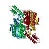
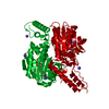
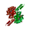
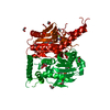
- PDB-6ly7: PylRS C-terminus domain mutant bound with 1-Formyl-L-tryptophan a... -
+ Open data

- Basic information
| Entry | Database: PDB / ID: 6ly7 | ||||||||||||
|---|---|---|---|---|---|---|---|---|---|---|---|---|---|
| Title | PylRS C-terminus domain mutant bound with 1-Formyl-L-tryptophan and AMPNP | ||||||||||||
 Components Components | Pyrrolysine--tRNA ligase | ||||||||||||
 Keywords Keywords | LIGASE / tRNA Synthetase | ||||||||||||
| Function / homology |  Function and homology information Function and homology informationpyrrolysine-tRNAPyl ligase / pyrrolysyl-tRNA synthetase activity / tRNA aminoacylation for protein translation / tRNA binding / ATP binding / cytoplasm Similarity search - Function | ||||||||||||
| Biological species |  Methanosarcina mazei (archaea) Methanosarcina mazei (archaea) | ||||||||||||
| Method |  X-RAY DIFFRACTION / SYNCHROTRON / MOLECULAR REPLACEMENT / Resolution: 2.09447266796 Å X-RAY DIFFRACTION / SYNCHROTRON / MOLECULAR REPLACEMENT / Resolution: 2.09447266796 Å | ||||||||||||
 Authors Authors | Weng, J.H. / Tsai, M.D. / Wang, Y.S. | ||||||||||||
| Funding support |  Taiwan, 3items Taiwan, 3items
| ||||||||||||
 Citation Citation | Journal: Biochemistry / Year: 2020 Title: Probing the Active Site of Deubiquitinase USP30 with Noncanonical Tryptophan Analogues. Authors: Jiang, H.K. / Wang, Y.H. / Weng, J.H. / Kurkute, P. / Li, C.L. / Lee, M.N. / Chen, P.J. / Tseng, H.W. / Tsai, M.D. / Wang, Y.S. | ||||||||||||
| History |
|
- Structure visualization
| Structure viewer | Molecule: MolmilJmol/JSmol |
|---|
- Downloads & links
-Download
| PDBx/mmCIF format | 6ly7.cif.gz | 129.8 KB | Display | PDBx/mmCIF format |
|---|---|---|---|---|
| PDB format | pdb6ly7.ent.gz | 91.6 KB | Display | PDB format |
| PDBx/mmJSON format | 6ly7.json.gz | Tree view | PDBx/mmJSON format | |
| Others |  Other downloads Other downloads |
-Validation report
| Arichive directory | https://data.pdbj.org/pub/pdb/validation_reports/ly/6ly7ftp://data.pdbj.org/pub/pdb/validation_reports/ly/6ly7 | HTTPS FTP |
|---|
-Related structure data
| Related structure data |  6ly3C 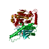 6ly6C 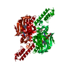 6lyaC  6lybC 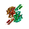 2zceS S: Starting model for refinement C: citing same article ( |
|---|---|
| Similar structure data |


-Links
 PDBj
PDBj


- Assembly
| Deposited unit | 
| ||||||||||||
|---|---|---|---|---|---|---|---|---|---|---|---|---|---|
| 1 | 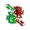
| ||||||||||||
| Unit cell |
|
-Components
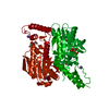
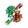
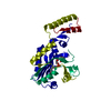
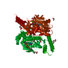
| #1: Protein | Mass: 31999.602 Da / Num. of mol.: 1 / Fragment: C-terminus domain / Mutation: N346G,C348Q,V401G Source method: isolated from a genetically manipulated source Source: (gene. exp.) Methanosarcina mazei (archaea) / Gene: pylS, DU43_20175, DU67_18120 / Production host:  References: UniProt: A0A0F8JXW8, UniProt: Q8PWY1*PLUS, pyrrolysine-tRNAPyl ligase | ||||||
|---|---|---|---|---|---|---|---|
| #2: Chemical | ChemComp-ANP /   Mass: 506.196 Da / Num. of mol.: 1 / Source method: obtained synthetically / Formula: C10H17N6O12P3 / Comment: AMP-PNP, energy-carrying molecule analogue*YM Mass: 506.196 Da / Num. of mol.: 1 / Source method: obtained synthetically / Formula: C10H17N6O12P3 / Comment: AMP-PNP, energy-carrying molecule analogue*YM | ||||||
| #3: Chemical |   Mass: 24.305 Da / Num. of mol.: 2 / Source method: obtained synthetically / Formula: Mg Mass: 24.305 Da / Num. of mol.: 2 / Source method: obtained synthetically / Formula: Mg#4: Chemical | ChemComp-TRF / |   Type: L-peptide linking / Mass: 232.235 Da / Num. of mol.: 1 / Source method: obtained synthetically / Formula: C12H12N2O3 / Feature type: SUBJECT OF INVESTIGATION Type: L-peptide linking / Mass: 232.235 Da / Num. of mol.: 1 / Source method: obtained synthetically / Formula: C12H12N2O3 / Feature type: SUBJECT OF INVESTIGATION#5: Water | ChemComp-HOH / |  Mass: 18.015 Da / Num. of mol.: 153 / Source method: isolated from a natural source / Formula: H2O Mass: 18.015 Da / Num. of mol.: 153 / Source method: isolated from a natural source / Formula: H2OHas ligand of interest | Y | |
-Experimental details
-Experiment
| Experiment | Method: X-RAY DIFFRACTION / Number of used crystals: 1 |
|---|
- Sample preparation
| Crystal | Density Matthews: 3.62 Å3/Da / Density % sol: 66.05 % |
|---|---|
| Crystal grow | Temperature: 293 K / Method: vapor diffusion, hanging drop / pH: 7.5 / Details: 100 mM HEPES, 10% PEG 8000, 10% Ethylene Glycerol |
-Data collection
| Diffraction | Mean temperature: 110 K / Serial crystal experiment: N |
|---|---|
| Diffraction source | Source: SYNCHROTRON / Site: NSRRC / Beamline: BL15A1 / Wavelength: 1 Å |
| Detector | Type: RAYONIX MX300HE / Detector: CCD / Date: Mar 27, 2017 |
| Radiation | Protocol: SINGLE WAVELENGTH / Monochromatic (M) / Laue (L): M / Scattering type: x-ray |
| Radiation wavelength | Wavelength: 1 Å / Relative weight: 1 |
| Reflection | Resolution: 2.09→28.3551 Å / Num. obs: 26941 / % possible obs: 96.89 % / Redundancy: 7.3 % / Biso Wilson estimate: 44.2768027561 Å2 / Rpim(I) all: 0.039 / Rrim(I) all: 0.104 / Net I/σ(I): 33.2 |
| Reflection shell | Resolution: 2.09→2.18 Å / Mean I/σ(I) obs: 2.5 / Num. unique obs: 1908 / Rpim(I) all: 0.308 / Rrim(I) all: 0.829 |
- Processing
| Software |
| |||||||||||||||||||||||||||||||||||||||||||||||||||||||||||||||||||||||||||||||||||||||||||||||||||||||||
|---|---|---|---|---|---|---|---|---|---|---|---|---|---|---|---|---|---|---|---|---|---|---|---|---|---|---|---|---|---|---|---|---|---|---|---|---|---|---|---|---|---|---|---|---|---|---|---|---|---|---|---|---|---|---|---|---|---|---|---|---|---|---|---|---|---|---|---|---|---|---|---|---|---|---|---|---|---|---|---|---|---|---|---|---|---|---|---|---|---|---|---|---|---|---|---|---|---|---|---|---|---|---|---|---|---|---|
| Refinement | Method to determine structure: MOLECULAR REPLACEMENT Starting model: 2ZCE Resolution: 2.09447266796→28.3550953944 Å / SU ML: 0.20472862989 / Cross valid method: FREE R-VALUE / σ(F): 1.35331964804 / Phase error: 19.5171703414 Stereochemistry target values: GeoStd + Monomer Library + CDL v1.2
| |||||||||||||||||||||||||||||||||||||||||||||||||||||||||||||||||||||||||||||||||||||||||||||||||||||||||
| Solvent computation | Shrinkage radii: 0.9 Å / VDW probe radii: 1.11 Å / Solvent model: FLAT BULK SOLVENT MODEL | |||||||||||||||||||||||||||||||||||||||||||||||||||||||||||||||||||||||||||||||||||||||||||||||||||||||||
| Displacement parameters | Biso mean: 51.4604466738 Å2 | |||||||||||||||||||||||||||||||||||||||||||||||||||||||||||||||||||||||||||||||||||||||||||||||||||||||||
| Refinement step | Cycle: LAST / Resolution: 2.09447266796→28.3550953944 Å
| |||||||||||||||||||||||||||||||||||||||||||||||||||||||||||||||||||||||||||||||||||||||||||||||||||||||||
| Refine LS restraints |
| |||||||||||||||||||||||||||||||||||||||||||||||||||||||||||||||||||||||||||||||||||||||||||||||||||||||||
| LS refinement shell |
|
