 Movie
Movie Controller

[English] 日本語
 Yorodumi
- PDB-1nvm: Crystal structure of a bifunctional aldolase-dehydrogenase : sequ... -
+ Open data

- Basic information
| Entry | Database: PDB / ID: 1nvm | ||||||
|---|---|---|---|---|---|---|---|
| Title | Crystal structure of a bifunctional aldolase-dehydrogenase : sequestering a reactive and volatile intermediate | ||||||
 Components Components |
| ||||||
 Keywords Keywords | LYASE/OXIDOREDUCTASE / Sequestered tunnel / substrate channeling / Bifunctional enzyme / LYASE-OXIDOREDUCTASE COMPLEX | ||||||
| Function / homology |  Function and homology information Function and homology information4-hydroxy-2-oxovalerate aldolase / 4-hydroxy-2-oxovalerate aldolase activity / phenol-containing compound catabolic process / 2-isopropylmalate synthase activity / acetaldehyde dehydrogenase (acetylating) / acetaldehyde dehydrogenase (acetylating) activity / : / L-leucine biosynthetic process / NAD binding / NADP binding / manganese ion binding Similarity search - Function | ||||||
| Biological species |  Pseudomonas sp. (bacteria) Pseudomonas sp. (bacteria) | ||||||
| Method |  X-RAY DIFFRACTION / SYNCHROTRON / MAD / Resolution: 1.7 Å X-RAY DIFFRACTION / SYNCHROTRON / MAD / Resolution: 1.7 Å | ||||||
 Authors Authors | Manjasetty, A.B. / Powlowski, J. / Vrielink, A. | ||||||
 Citation Citation | Journal: Proc.Natl.Acad.Sci.USA / Year: 2003 Title: Crystal structure of a bifunctional aldolase-dehydrogenase: Sequestering a reactive and volatile intermediate Authors: Manjasetty, A.B. / Powlowski, J. / Vrielink, A. #1: Journal: Acta Crystallogr.,Sect.D / Year: 2001Title: Crystallization and preliminary X-ray analysis of dmpFG-encoded 4-hydroxy-2-ketovalerate aldolase-aldehyde dehydrogenase (acylating) from Pseudomonas sp.strain CF600 | ||||||
| History |
|
- Structure visualization
| Structure viewer | Molecule: MolmilJmol/JSmol |
|---|
- Downloads & links
-Download
| PDBx/mmCIF format | 1nvm.cif.gz | 559.1 KB | Display | PDBx/mmCIF format |
|---|---|---|---|---|
| PDB format | pdb1nvm.ent.gz | 451 KB | Display | PDB format |
| PDBx/mmJSON format | 1nvm.json.gz | Tree view | PDBx/mmJSON format | |
| Others |  Other downloads Other downloads |
-Validation report
| Arichive directory | https://data.pdbj.org/pub/pdb/validation_reports/nv/1nvmftp://data.pdbj.org/pub/pdb/validation_reports/nv/1nvm | HTTPS FTP |
|---|
-Related structure data
| Similar structure data |
|---|


-Links
 PDBj
PDBj- Assembly
| Deposited unit | 
| ||||||||
|---|---|---|---|---|---|---|---|---|---|
| 1 | 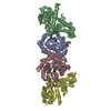
| ||||||||
| 2 | 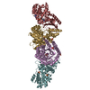
| ||||||||
| 3 | 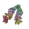
| ||||||||
| Unit cell |
|
-Components
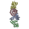
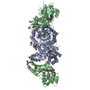
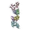
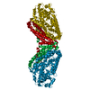
-Protein , 2 types, 8 molecules ACEGBDFH
| #1: Protein | Mass: 37520.645 Da / Num. of mol.: 4 Source method: isolated from a genetically manipulated source Source: (gene. exp.) Pseudomonas sp. (bacteria) / Strain: CF600 / Gene: DMPG / Plasmid: pT7-5 / Production host: References: UniProt: P51016, Lyases; Carbon-carbon lyases; Oxo-acid-lyases #2: Protein | Mass: 32713.812 Da / Num. of mol.: 4 Source method: isolated from a genetically manipulated source Source: (gene. exp.) Pseudomonas sp. (bacteria) / Strain: CF600 / Gene: DMPF / Plasmid: pT7-5 / Production host: References: UniProt: Q52060, acetaldehyde dehydrogenase (acetylating) |
|---|
-Non-polymers , 6 types, 2840 molecules 
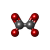


| #3: Chemical | ChemComp-MN /  Mass: 54.938 Da / Num. of mol.: 4 / Source method: obtained synthetically / Formula: Mn Mass: 54.938 Da / Num. of mol.: 4 / Source method: obtained synthetically / Formula: Mn#4: Chemical | ChemComp-OXL /  Mass: 88.019 Da / Num. of mol.: 4 / Source method: obtained synthetically / Formula: C2O4 Mass: 88.019 Da / Num. of mol.: 4 / Source method: obtained synthetically / Formula: C2O4#5: Chemical | ChemComp-SO4 /  Mass: 96.063 Da / Num. of mol.: 12 / Source method: obtained synthetically / Formula: SO4 Mass: 96.063 Da / Num. of mol.: 12 / Source method: obtained synthetically / Formula: SO4#6: Chemical | ChemComp-MPD / (  Mass: 118.174 Da / Num. of mol.: 6 / Source method: obtained synthetically / Formula: C6H14O2 / Comment: precipitant*YM Mass: 118.174 Da / Num. of mol.: 6 / Source method: obtained synthetically / Formula: C6H14O2 / Comment: precipitant*YM#7: Chemical |  Mass: 663.425 Da / Num. of mol.: 3 / Source method: obtained synthetically / Formula: C21H27N7O14P2 / Comment: NAD*YM Mass: 663.425 Da / Num. of mol.: 3 / Source method: obtained synthetically / Formula: C21H27N7O14P2 / Comment: NAD*YM#8: Water | ChemComp-HOH / | Mass: 18.015 Da / Num. of mol.: 2811 / Source method: isolated from a natural source / Formula: H2O |
|---|
-Experimental details
-Experiment
| Experiment | Method: X-RAY DIFFRACTION / Number of used crystals: 1 |
|---|
- Sample preparation
| Crystal | Density Matthews: 2.16 Å3/Da / Density % sol: 42.73 % / Description: 112 SE SUBSTRUCTURE | ||||||||||||||||||||||||||||||||||||||||||
|---|---|---|---|---|---|---|---|---|---|---|---|---|---|---|---|---|---|---|---|---|---|---|---|---|---|---|---|---|---|---|---|---|---|---|---|---|---|---|---|---|---|---|---|
| Crystal grow | Temperature: 310 K / pH: 7.5 Details: PEG 8000, Ammonium sulfate, PIPES, pH 7.5, Hanging drop and micro seeding, temperature 310.0K, pH 7.50 | ||||||||||||||||||||||||||||||||||||||||||
| Crystal grow | *PLUS Temperature: 290 K / pH: 7.4 / Method: vapor diffusion, hanging dropDetails: Manjasetty, B.A., (2001) Acta Crystallogr.,Sect.D, 57, 582. | ||||||||||||||||||||||||||||||||||||||||||
| Components of the solutions | *PLUS
|
-Data collection
| Diffraction | Mean temperature: 100 K |
|---|---|
| Diffraction source | Source: SYNCHROTRON / Site: ALS  / Beamline: 5.0.2 / Wavelength: 1 / Beamline: 5.0.2 / Wavelength: 1 |
| Detector | Type: ADSC QUANTUM 4 / Detector: CCD / Date: Dec 14, 2000 / Details: MIRRORS |
| Radiation | Monochromator: CURVED-CRYSTAL SI 111 / Protocol: MULTIPLE WAVELENGTH / Monochromatic (M) / Laue (L): M / Scattering type: x-ray |
| Radiation wavelength | Wavelength: 1 Å / Relative weight: 1 |
| Reflection | Resolution: 1.7→100 Å / Num. obs: 299052 / % possible obs: 99.7 % / Observed criterion σ(I): 0 / Redundancy: 6.2 % / Biso Wilson estimate: 28.3 Å2 / Rmerge(I) obs: 0.078 / Rsym value: 0.078 / Net I/σ(I): 18.7 |
| Reflection shell | Resolution: 1.7→1.74 Å / Redundancy: 4 % / Rmerge(I) obs: 0.565 / Mean I/σ(I) obs: 2 / Rsym value: 0.565 / % possible all: 99.3 |
| Reflection | *PLUS Highest resolution: 1.7 Å / Num. measured all: 1829280 |
| Reflection shell | *PLUS % possible obs: 99.3 % / Mean I/σ(I) obs: 2 |
- Processing
| Software |
| ||||||||||||||||||||||||||||||||||||||||||||||||||||||||||||||||||||||||||||||||||||
|---|---|---|---|---|---|---|---|---|---|---|---|---|---|---|---|---|---|---|---|---|---|---|---|---|---|---|---|---|---|---|---|---|---|---|---|---|---|---|---|---|---|---|---|---|---|---|---|---|---|---|---|---|---|---|---|---|---|---|---|---|---|---|---|---|---|---|---|---|---|---|---|---|---|---|---|---|---|---|---|---|---|---|---|---|---|
| Refinement | Method to determine structure: MAD / Resolution: 1.7→20 Å / Isotropic thermal model: Isotropic / σ(F): 0 / Stereochemistry target values: Engh & Huber / Details: MAXIMUM LIKLIHOOD METHOD
| ||||||||||||||||||||||||||||||||||||||||||||||||||||||||||||||||||||||||||||||||||||
| Displacement parameters | Biso mean: 28.5 Å2 | ||||||||||||||||||||||||||||||||||||||||||||||||||||||||||||||||||||||||||||||||||||
| Refinement step | Cycle: LAST / Resolution: 1.7→20 Å
| ||||||||||||||||||||||||||||||||||||||||||||||||||||||||||||||||||||||||||||||||||||
| Refine LS restraints |
| ||||||||||||||||||||||||||||||||||||||||||||||||||||||||||||||||||||||||||||||||||||
| LS refinement shell | Resolution: 1.7→1.783 Å /
| ||||||||||||||||||||||||||||||||||||||||||||||||||||||||||||||||||||||||||||||||||||
| Refinement | *PLUS Highest resolution: 1.7 Å / Lowest resolution: 20 Å / Rfactor Rfree: 0.232 | ||||||||||||||||||||||||||||||||||||||||||||||||||||||||||||||||||||||||||||||||||||
| Solvent computation | *PLUS | ||||||||||||||||||||||||||||||||||||||||||||||||||||||||||||||||||||||||||||||||||||
| Displacement parameters | *PLUS | ||||||||||||||||||||||||||||||||||||||||||||||||||||||||||||||||||||||||||||||||||||
| Refine LS restraints | *PLUS
|
