 Movie
Movie Controller

[English] 日本語
 Yorodumi
- PDB-4lrs: Crystal and solution structures of the bifunctional enzyme (Aldol... -
+ Open data

- Basic information
| Entry | Database: PDB / ID: 4lrs | |||||||||
|---|---|---|---|---|---|---|---|---|---|---|
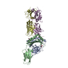
| Title | Crystal and solution structures of the bifunctional enzyme (Aldolase/Aldehyde dehydrogenase) from Thermomonospora curvata, reveal a cofactor-binding domain motion during NAD+ and CoA accommodation whithin the shared cofactor-binding site | |||||||||
 Components Components |
| |||||||||
 Keywords Keywords | OXIDOREDUCTASE / Rossmann fold / TIM barrel domain / Dehydrogenase / aldolase | |||||||||
| Function / homology |  Function and homology information Function and homology information4-hydroxy-2-oxovalerate aldolase / 4-hydroxy-2-oxovalerate aldolase activity / 2-isopropylmalate synthase activity / acetaldehyde dehydrogenase (acetylating) / acetaldehyde dehydrogenase (acetylating) activity / L-leucine biosynthetic process / NAD binding / manganese ion binding Similarity search - Function | |||||||||
| Biological species |  Thermomonospora curvata (bacteria) Thermomonospora curvata (bacteria) | |||||||||
| Method |  X-RAY DIFFRACTION / SYNCHROTRON / MOLECULAR REPLACEMENT / Resolution: 1.55 Å X-RAY DIFFRACTION / SYNCHROTRON / MOLECULAR REPLACEMENT / Resolution: 1.55 Å | |||||||||
 Authors Authors | Fischer, B. / Branlant, G. / Talfournier, F. / Gruez, A. | |||||||||
 Citation Citation | Journal: To be Published Title: Crystal and solution structures of the bifunctional enzyme (Aldolase/Aldehyde dehydrogenase) from Thermomonospora curvata, reveal a cofactor-binding domain motion during NAD+ and CoA ...Title: Crystal and solution structures of the bifunctional enzyme (Aldolase/Aldehyde dehydrogenase) from Thermomonospora curvata, reveal a cofactor-binding domain motion during NAD+ and CoA accommodation whithin the shared cofactor-binding site Authors: Fischer, B. / Branlant, G. / Talfournier, F. / Gruez, A. | |||||||||
| History |
|
- Structure visualization
| Structure viewer | Molecule: MolmilJmol/JSmol |
|---|
- Downloads & links
-Download
| PDBx/mmCIF format | 4lrs.cif.gz | 287.9 KB | Display | PDBx/mmCIF format |
|---|---|---|---|---|
| PDB format | pdb4lrs.ent.gz | 231.2 KB | Display | PDB format |
| PDBx/mmJSON format | 4lrs.json.gz | Tree view | PDBx/mmJSON format | |
| Others |  Other downloads Other downloads |
-Validation report
| Arichive directory | https://data.pdbj.org/pub/pdb/validation_reports/lr/4lrsftp://data.pdbj.org/pub/pdb/validation_reports/lr/4lrs | HTTPS FTP |
|---|
-Related structure data
| Related structure data |  4lrtC  1nvmS C: citing same article ( S: Starting model for refinement |
|---|---|
| Similar structure data |


-Links
 PDBj
PDBj

- Assembly
| Deposited unit | 
| ||||||||
|---|---|---|---|---|---|---|---|---|---|
| 1 | 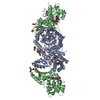
| ||||||||
| 2 | 
| ||||||||
| Unit cell |
|
-Components
-Protein , 2 types, 2 molecules AB
| #1: Protein | Mass: 37326.852 Da / Num. of mol.: 1 Source method: isolated from a genetically manipulated source Source: (gene. exp.) Thermomonospora curvata (bacteria) / Strain: DSM 43183 / Gene: Tcur_0536 / Plasmid: pET28b / Production host: References: UniProt: D1A3K8, 4-hydroxy-2-oxovalerate aldolase |
|---|---|
| #2: Protein | Mass: 34127.828 Da / Num. of mol.: 1 Source method: isolated from a genetically manipulated source Source: (gene. exp.) Thermomonospora curvata (bacteria) / Strain: DSM 43183 / Gene: Tcur_0535 / Plasmid: pET28b / Production host: References: UniProt: D1A3K7, acetaldehyde dehydrogenase (acetylating) |
-Protein/peptide , 1 types, 1 molecules N
| #3: Protein/peptide | Mass: 231.249 Da / Num. of mol.: 1 Source method: isolated from a genetically manipulated source Source: (gene. exp.) Thermomonospora curvata (bacteria) / Strain: DSM 43183 / Plasmid: pET28b / Production host: |
|---|
-Non-polymers , 9 types, 585 molecules 


| #4: Chemical | ChemComp-SO4 /  Mass: 96.063 Da / Num. of mol.: 1 / Source method: obtained synthetically / Formula: SO4 Mass: 96.063 Da / Num. of mol.: 1 / Source method: obtained synthetically / Formula: SO4 | ||||||||||||
|---|---|---|---|---|---|---|---|---|---|---|---|---|---|
| #5: Chemical | ChemComp-PYR /  Mass: 88.062 Da / Num. of mol.: 1 / Source method: obtained synthetically / Formula: C3H4O3 Mass: 88.062 Da / Num. of mol.: 1 / Source method: obtained synthetically / Formula: C3H4O3 | ||||||||||||
| #6: Chemical |  Mass: 35.453 Da / Num. of mol.: 2 / Source method: obtained synthetically / Formula: Cl Mass: 35.453 Da / Num. of mol.: 2 / Source method: obtained synthetically / Formula: Cl#7: Chemical | ChemComp-MG / |  Mass: 24.305 Da / Num. of mol.: 1 / Source method: obtained synthetically / Formula: Mg Mass: 24.305 Da / Num. of mol.: 1 / Source method: obtained synthetically / Formula: Mg#8: Chemical | ChemComp-GOL /  Mass: 92.094 Da / Num. of mol.: 6 / Source method: obtained synthetically / Formula: C3H8O3 Mass: 92.094 Da / Num. of mol.: 6 / Source method: obtained synthetically / Formula: C3H8O3#9: Chemical | ChemComp-PEG /  Mass: 106.120 Da / Num. of mol.: 7 / Source method: obtained synthetically / Formula: C4H10O3 Mass: 106.120 Da / Num. of mol.: 7 / Source method: obtained synthetically / Formula: C4H10O3#10: Chemical | ChemComp-NAD / |  Mass: 663.425 Da / Num. of mol.: 1 / Source method: obtained synthetically / Formula: C21H27N7O14P2 / Comment: NAD*YM Mass: 663.425 Da / Num. of mol.: 1 / Source method: obtained synthetically / Formula: C21H27N7O14P2 / Comment: NAD*YM#11: Chemical | ChemComp-FOR / |  Mass: 30.026 Da / Num. of mol.: 1 / Source method: obtained synthetically / Formula: CH2O Mass: 30.026 Da / Num. of mol.: 1 / Source method: obtained synthetically / Formula: CH2O#12: Water | ChemComp-HOH / | Mass: 18.015 Da / Num. of mol.: 565 / Source method: isolated from a natural source / Formula: H2O |
-Experimental details
-Experiment
| Experiment | Method: X-RAY DIFFRACTION |
|---|
- Sample preparation
| Crystal | Density Matthews: 2.68 Å3/Da / Density % sol: 54.03 % |
|---|---|
| Crystal grow | Temperature: 298 K / Method: vapor diffusion, hanging drop Details: 26-34% PEG 4000 or 3350, 0.1 M Tris, 0.2 M Li2SO4, 0.005M NAD, VAPOR DIFFUSION, HANGING DROP, temperature 298.0K PH range: 7.5-9.0 |
-Data collection
| Diffraction | Mean temperature: 100 K |
|---|---|
| Diffraction source | Source: SYNCHROTRON / Site: SOLEIL  / Beamline: PROXIMA 1 / Wavelength: 0.8856 / Beamline: PROXIMA 1 / Wavelength: 0.8856 |
| Detector | Type: ADSC QUANTUM 315r / Detector: CCD / Date: Apr 11, 2011 |
| Radiation | Monochromator: KIRKPATRICK-BAEZ PAIR OF BI- MORPH MIRRORS PLUS CHANNEL CUT CRYOGENICALLY COOLED MONOCHROMATOR CRYSTAL Protocol: SINGLE WAVELENGTH / Monochromatic (M) / Laue (L): M / Scattering type: x-ray |
| Radiation wavelength | Wavelength: 0.8856 Å / Relative weight: 1 |
| Reflection | Resolution: 1.55→43.35 Å / Num. obs: 424737 / % possible obs: 97.6 % / Observed criterion σ(I): 3 / Redundancy: 3.98 % / Rmerge(I) obs: 0.059 / Net I/σ(I): 13.68 |
| Reflection shell | Resolution: 1.55→1.59 Å / Redundancy: 3.01 % / Rmerge(I) obs: 0.365 / Mean I/σ(I) obs: 3.06 / % possible all: 85.2 |
- Processing
| Software |
| ||||||||||||||||||||||||||||||||||||||||||||||||||||||||||||||||||||||||||||||||||||||||||||||||||||||||||||||||||||||||||||||||||||||||||||||||||||||||||||||||||||||||||||||||||||||
|---|---|---|---|---|---|---|---|---|---|---|---|---|---|---|---|---|---|---|---|---|---|---|---|---|---|---|---|---|---|---|---|---|---|---|---|---|---|---|---|---|---|---|---|---|---|---|---|---|---|---|---|---|---|---|---|---|---|---|---|---|---|---|---|---|---|---|---|---|---|---|---|---|---|---|---|---|---|---|---|---|---|---|---|---|---|---|---|---|---|---|---|---|---|---|---|---|---|---|---|---|---|---|---|---|---|---|---|---|---|---|---|---|---|---|---|---|---|---|---|---|---|---|---|---|---|---|---|---|---|---|---|---|---|---|---|---|---|---|---|---|---|---|---|---|---|---|---|---|---|---|---|---|---|---|---|---|---|---|---|---|---|---|---|---|---|---|---|---|---|---|---|---|---|---|---|---|---|---|---|---|---|---|---|
| Refinement | Method to determine structure: MOLECULAR REPLACEMENT Starting model: PDB ENTRY 1NVM Resolution: 1.55→25 Å / Cor.coef. Fo:Fc: 0.971 / Cor.coef. Fo:Fc free: 0.957 / SU B: 3.01 / SU ML: 0.049 / Cross valid method: THROUGHOUT / σ(F): 3 / ESU R: 0.09 / ESU R Free: 0.076 / Stereochemistry target values: MAXIMUM LIKELIHOOD / Details: HYDROGENS HAVE BEEN USED IF PRESENT IN THE INPUT
| ||||||||||||||||||||||||||||||||||||||||||||||||||||||||||||||||||||||||||||||||||||||||||||||||||||||||||||||||||||||||||||||||||||||||||||||||||||||||||||||||||||||||||||||||||||||
| Solvent computation | Ion probe radii: 0.8 Å / Shrinkage radii: 0.8 Å / VDW probe radii: 1.2 Å / Solvent model: MASK | ||||||||||||||||||||||||||||||||||||||||||||||||||||||||||||||||||||||||||||||||||||||||||||||||||||||||||||||||||||||||||||||||||||||||||||||||||||||||||||||||||||||||||||||||||||||
| Displacement parameters | Biso mean: 12.42 Å2
| ||||||||||||||||||||||||||||||||||||||||||||||||||||||||||||||||||||||||||||||||||||||||||||||||||||||||||||||||||||||||||||||||||||||||||||||||||||||||||||||||||||||||||||||||||||||
| Refinement step | Cycle: LAST / Resolution: 1.55→25 Å
| ||||||||||||||||||||||||||||||||||||||||||||||||||||||||||||||||||||||||||||||||||||||||||||||||||||||||||||||||||||||||||||||||||||||||||||||||||||||||||||||||||||||||||||||||||||||
| Refine LS restraints |
|