 Movie
Movie Controller

[English] 日本語
 Yorodumi
- PDB-4xi5: gHgL of varicella-zoster virus in complex with human neutralizing... -
+ Open data

- Basic information
| Entry | Database: PDB / ID: 4xi5 | |||||||||
|---|---|---|---|---|---|---|---|---|---|---|
| Title | gHgL of varicella-zoster virus in complex with human neutralizing antibodies | |||||||||
 Components Components |
| |||||||||
 Keywords Keywords | Viral Protein/Immune System / complex / neutralization epitope / Viral Protein-Immune System complex | |||||||||
| Function / homology |  Function and homology information Function and homology informationhost cell endosome membrane / host cell Golgi apparatus / fusion of virus membrane with host plasma membrane / viral envelope / symbiont entry into host cell / host cell plasma membrane / virion membrane Similarity search - Function | |||||||||
| Biological species |  Human herpesvirus 3 strain Oka vaccine Human herpesvirus 3 strain Oka vaccine Homo sapiens (human) Homo sapiens (human) | |||||||||
| Method |  X-RAY DIFFRACTION / SYNCHROTRON / MOLECULAR REPLACEMENT / Resolution: 3.9 Å X-RAY DIFFRACTION / SYNCHROTRON / MOLECULAR REPLACEMENT / Resolution: 3.9 Å | |||||||||
 Authors Authors | Xing, Y. | |||||||||
 Citation Citation | Journal: Proc.Natl.Acad.Sci.USA / Year: 2015 Title: A site of varicella-zoster virus vulnerability identified by structural studies of neutralizing antibodies bound to the glycoprotein complex gHgL. Authors: Xing, Y. / Oliver, S.L. / Nguyen, T. / Ciferri, C. / Nandi, A. / Hickman, J. / Giovani, C. / Yang, E. / Palladino, G. / Grose, C. / Uematsu, Y. / Lilja, A.E. / Arvin, A.M. / Carfi, A. | |||||||||
| History |
|
- Structure visualization
| Structure viewer | Molecule: MolmilJmol/JSmol |
|---|
- Downloads & links
-Download
| PDBx/mmCIF format | 4xi5.cif.gz | 264.4 KB | Display | PDBx/mmCIF format |
|---|---|---|---|---|
| PDB format | pdb4xi5.ent.gz | 204.8 KB | Display | PDB format |
| PDBx/mmJSON format | 4xi5.json.gz | Tree view | PDBx/mmJSON format | |
| Others |  Other downloads Other downloads |
-Validation report
| Arichive directory | https://data.pdbj.org/pub/pdb/validation_reports/xi/4xi5ftp://data.pdbj.org/pub/pdb/validation_reports/xi/4xi5 | HTTPS FTP |
|---|
-Related structure data
| Related structure data |  4xhjSC S: Starting model for refinement C: citing same article ( |
|---|---|
| Similar structure data |


-Links
 PDBj
PDBj

- Assembly
| Deposited unit | 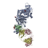
| ||||||||
|---|---|---|---|---|---|---|---|---|---|
| 1 |
| ||||||||
| Unit cell |
|
-Components
-Envelope glycoprotein ... , 2 types, 2 molecules AB
| #1: Protein | Mass: 92763.609 Da / Num. of mol.: 1 Source method: isolated from a genetically manipulated source Source: (gene. exp.) Human herpesvirus 3 strain Oka vaccine / Gene: gH, ORF37 / Production host: Homo sapiens (human) / References: UniProt: Q775J3 |
|---|---|
| #2: Protein | Mass: 15197.178 Da / Num. of mol.: 1 Source method: isolated from a genetically manipulated source Source: (gene. exp.) Human herpesvirus 3 strain Oka vaccine / Gene: gL, ORF60 / Production host: Homo sapiens (human) / References: UniProt: Q9J3N1 |
-Antibody , 2 types, 2 molecules CD
| #3: Antibody | Mass: 23172.715 Da / Num. of mol.: 1 Source method: isolated from a genetically manipulated source Source: (gene. exp.) Homo sapiens (human) / Production host: Homo sapiens (human) |
|---|---|
| #4: Antibody | Mass: 30198.682 Da / Num. of mol.: 1 Source method: isolated from a genetically manipulated source Source: (gene. exp.) Homo sapiens (human) / Production host: Homo sapiens (human) |
-Sugars , 2 types, 4 molecules 
| #5: Polysaccharide | alpha-D-mannopyranose-(1-3)-beta-D-mannopyranose-(1-4)-2-acetamido-2-deoxy-beta-D-glucopyranose-(1- ...alpha-D-mannopyranose-(1-3)-beta-D-mannopyranose-(1-4)-2-acetamido-2-deoxy-beta-D-glucopyranose-(1-4)-2-acetamido-2-deoxy-beta-D-glucopyranose |
|---|---|
| #6: Sugar |  Type: D-saccharide, beta linking / Mass: 221.208 Da / Num. of mol.: 3 / Source method: obtained synthetically / Formula: C8H15NO6 Type: D-saccharide, beta linking / Mass: 221.208 Da / Num. of mol.: 3 / Source method: obtained synthetically / Formula: C8H15NO6 |
-Details
| Has protein modification | Y |
|---|
-Experimental details
-Experiment
| Experiment | Method: X-RAY DIFFRACTION |
|---|
- Sample preparation
| Crystal | Density Matthews: 4.2 Å3/Da / Density % sol: 70.68 % |
|---|---|
| Crystal grow | Temperature: 293 K / Method: vapor diffusion, hanging drop / pH: 6 / Details: 0.1M MES pH 6.0 and 4% PEG 4000 |
-Data collection
| Diffraction | Mean temperature: 100 K |
|---|---|
| Diffraction source | Source: SYNCHROTRON / Site: APS  / Beamline: 17-ID / Wavelength: 1 Å / Beamline: 17-ID / Wavelength: 1 Å |
| Detector | Type: DECTRIS PILATUS 6M / Detector: PIXEL / Date: Jun 21, 2013 |
| Radiation | Protocol: SINGLE WAVELENGTH / Monochromatic (M) / Laue (L): M / Scattering type: x-ray |
| Radiation wavelength | Wavelength: 1 Å / Relative weight: 1 |
| Reflection | Resolution: 3.8852→200 Å / Num. obs: 23170 / % possible obs: 99.1 % / Redundancy: 6.3 % / Rmerge(I) obs: 0.067 / Net I/σ(I): 24.4 |
| Reflection shell | Resolution: 3.8852→3.97 Å / Redundancy: 5.4 % / Rmerge(I) obs: 0.753 / Mean I/σ(I) obs: 1.7 |
- Processing
| Software |
| |||||||||||||||||||||||||||||||||||||||||||||||||||||||||||||||
|---|---|---|---|---|---|---|---|---|---|---|---|---|---|---|---|---|---|---|---|---|---|---|---|---|---|---|---|---|---|---|---|---|---|---|---|---|---|---|---|---|---|---|---|---|---|---|---|---|---|---|---|---|---|---|---|---|---|---|---|---|---|---|---|---|
| Refinement | Method to determine structure: MOLECULAR REPLACEMENT Starting model: PDB entry 4XHJ Resolution: 3.9→37.322 Å / SU ML: 0.45 / Cross valid method: FREE R-VALUE / σ(F): 1.35 / Phase error: 32.36 / Stereochemistry target values: ML
| |||||||||||||||||||||||||||||||||||||||||||||||||||||||||||||||
| Solvent computation | Shrinkage radii: 0.9 Å / VDW probe radii: 1.11 Å / Solvent model: FLAT BULK SOLVENT MODEL | |||||||||||||||||||||||||||||||||||||||||||||||||||||||||||||||
| Refinement step | Cycle: LAST / Resolution: 3.9→37.322 Å
| |||||||||||||||||||||||||||||||||||||||||||||||||||||||||||||||
| Refine LS restraints |
| |||||||||||||||||||||||||||||||||||||||||||||||||||||||||||||||
| LS refinement shell |
|
