 Movie
Movie Controller


+ Open data

- Basic information
| Entry | Database: PDB / ID: 4gsk | ||||||
|---|---|---|---|---|---|---|---|
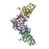
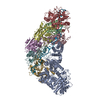
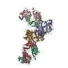
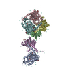
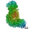
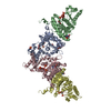
| Title | Crystal structure of an Atg7-Atg10 crosslinked complex | ||||||
 Components Components |
| ||||||
 Keywords Keywords | PROTEIN TRANSPORT/LIGASE / Ubiquitin-like protein activation enzyme / Ubiquitin-like protein transfer enzyme / PROTEIN TRANSPORT-LIGASE complex | ||||||
| Function / homology |  Function and homology information Function and homology informationAtg12 transferase activity / Atg12 activating enzyme activity / Atg8 activating enzyme activity / protein modification by small protein conjugation / extrinsic component of phagophore assembly site membrane / Macroautophagy / cytoplasm to vacuole targeting by the Cvt pathway / nucleophagy / phagophore assembly site membrane / autophagy of mitochondrion ...Atg12 transferase activity / Atg12 activating enzyme activity / Atg8 activating enzyme activity / protein modification by small protein conjugation / extrinsic component of phagophore assembly site membrane / Macroautophagy / cytoplasm to vacuole targeting by the Cvt pathway / nucleophagy / phagophore assembly site membrane / autophagy of mitochondrion / piecemeal microautophagy of the nucleus / phagophore assembly site / Transferases; Acyltransferases; Aminoacyltransferases / cellular response to nitrogen starvation / Antigen processing: Ubiquitination & Proteasome degradation / autophagosome assembly / mitophagy / Neutrophil degranulation / macroautophagy / autophagy / mitochondrion / membrane / identical protein binding / cytoplasm / cytosol Similarity search - Function | ||||||
| Biological species |  | ||||||
| Method |  X-RAY DIFFRACTION / SYNCHROTRON / MOLECULAR REPLACEMENT / Resolution: 2.9 Å X-RAY DIFFRACTION / SYNCHROTRON / MOLECULAR REPLACEMENT / Resolution: 2.9 Å | ||||||
 Authors Authors | Kaiser, S.E. / Mao, K. / Taherbhoy, A.M. / Yu, S. / Olszewski, J.L. / Duda, D.M. / Kurinov, I. / Deng, A. / Fenn, T.D. / Klionsky, D.J. / Schulman, B.A. | ||||||
 Citation Citation | Journal: Nat.Struct.Mol.Biol. / Year: 2012 Title: Noncanonical E2 recruitment by the autophagy E1 revealed by Atg7-Atg3 and Atg7-Atg10 structures. Authors: Kaiser, S.E. / Mao, K. / Taherbhoy, A.M. / Yu, S. / Olszewski, J.L. / Duda, D.M. / Kurinov, I. / Deng, A. / Fenn, T.D. / Klionsky, D.J. / Schulman, B.A. | ||||||
| History |
|
- Structure visualization
| Structure viewer | Molecule: MolmilJmol/JSmol |
|---|
- Downloads & links
-Download
| PDBx/mmCIF format | 4gsk.cif.gz | 287.5 KB | Display | PDBx/mmCIF format |
|---|---|---|---|---|
| PDB format | pdb4gsk.ent.gz | 230.8 KB | Display | PDB format |
| PDBx/mmJSON format | 4gsk.json.gz | Tree view | PDBx/mmJSON format | |
| Others |  Other downloads Other downloads |
-Validation report
| Arichive directory | https://data.pdbj.org/pub/pdb/validation_reports/gs/4gskftp://data.pdbj.org/pub/pdb/validation_reports/gs/4gsk | HTTPS FTP |
|---|
-Related structure data
| Related structure data |  4gsjC  4gslC  3t7eS  3t7fS C: citing same article ( S: Starting model for refinement |
|---|---|
| Similar structure data |


-Links
 PDBj
PDBj


- Assembly
| Deposited unit | 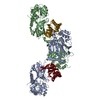
| ||||||||
|---|---|---|---|---|---|---|---|---|---|
| 1 |
| ||||||||
| Unit cell |
|
-Components
| #1: Protein | Mass: 69628.016 Da / Num. of mol.: 2 / Fragment: UNP residues 1-613 / Mutation: C39S, C195S, C375A Source method: isolated from a genetically manipulated source Source: (gene. exp.) Strain: ATCC 204508 / S288c / Gene: APG7, ATG7, CVT2, YHR171W / Plasmid: pGEX-4T1 / Production host:  #2: Protein | Mass: 20147.807 Da / Num. of mol.: 2 / Mutation: C26S, C137S Source method: isolated from a genetically manipulated source Source: (gene. exp.) Strain: ATCC 204508 / S288c / Gene: APG10, ATG10, YLL042C / Plasmid: pGEX-4T1 / Production host: References: UniProt: Q07879, Ligases; Forming carbon-nitrogen bonds; Acid-amino-acid ligases (peptide synthases) #3: Chemical |   Mass: 65.409 Da / Num. of mol.: 2 / Source method: obtained synthetically / Formula: Zn Mass: 65.409 Da / Num. of mol.: 2 / Source method: obtained synthetically / Formula: Zn |
|---|
-Experimental details
-Experiment
| Experiment | Method: X-RAY DIFFRACTION / Number of used crystals: 1 |
|---|
- Sample preparation
| Crystal | Density Matthews: 2.66 Å3/Da / Density % sol: 53.78 % |
|---|---|
| Crystal grow | Temperature: 294 K / Method: vapor diffusion, sitting drop / pH: 6.7 Details: 76.5 mM sodium/potassium phosphate, pH 6.5, 9 mM Tris, pH 8.5, 153 mM sodium chloride, 100 mM glycine, 72 mM sodium/potassium tartrate, 19.125% PEG1000, 0.045% PEG5000 MME, 4.5% dioxane, ...Details: 76.5 mM sodium/potassium phosphate, pH 6.5, 9 mM Tris, pH 8.5, 153 mM sodium chloride, 100 mM glycine, 72 mM sodium/potassium tartrate, 19.125% PEG1000, 0.045% PEG5000 MME, 4.5% dioxane, VAPOR DIFFUSION, SITTING DROP, temperature 294K |
-Data collection
| Diffraction | Mean temperature: 100 K | |||||||||||||||||||||||||||||||||||||||||||||||||||||||||||||||||||||||||||||
|---|---|---|---|---|---|---|---|---|---|---|---|---|---|---|---|---|---|---|---|---|---|---|---|---|---|---|---|---|---|---|---|---|---|---|---|---|---|---|---|---|---|---|---|---|---|---|---|---|---|---|---|---|---|---|---|---|---|---|---|---|---|---|---|---|---|---|---|---|---|---|---|---|---|---|---|---|---|---|
| Diffraction source | Source: SYNCHROTRON / Site: APS  / Beamline: 24-ID-C / Wavelength: 0.9795 Å / Beamline: 24-ID-C / Wavelength: 0.9795 Å | |||||||||||||||||||||||||||||||||||||||||||||||||||||||||||||||||||||||||||||
| Detector | Type: ADSC QUANTUM 315 / Detector: CCD / Date: Mar 29, 2012 | |||||||||||||||||||||||||||||||||||||||||||||||||||||||||||||||||||||||||||||
| Radiation | Monochromator: double crystal Si(111) / Protocol: SINGLE WAVELENGTH / Monochromatic (M) / Laue (L): M / Scattering type: x-ray | |||||||||||||||||||||||||||||||||||||||||||||||||||||||||||||||||||||||||||||
| Radiation wavelength | Wavelength: 0.9795 Å / Relative weight: 1 | |||||||||||||||||||||||||||||||||||||||||||||||||||||||||||||||||||||||||||||
| Reflection | Resolution: 2.9→50 Å / Num. all: 43151 / Num. obs: 42754 / % possible obs: 99.1 % / Observed criterion σ(F): 0 / Observed criterion σ(I): 0 / Redundancy: 3.2 % / Rmerge(I) obs: 0.134 / Χ2: 1.083 / Net I/σ(I): 9.9 | |||||||||||||||||||||||||||||||||||||||||||||||||||||||||||||||||||||||||||||
| Reflection shell |
|
- Processing
| Software |
| ||||||||||||||||||||||||||||||||||||||||||||||||||||||||||||||||||||||||||||||||||||||||||||||||||||||||||||||||
|---|---|---|---|---|---|---|---|---|---|---|---|---|---|---|---|---|---|---|---|---|---|---|---|---|---|---|---|---|---|---|---|---|---|---|---|---|---|---|---|---|---|---|---|---|---|---|---|---|---|---|---|---|---|---|---|---|---|---|---|---|---|---|---|---|---|---|---|---|---|---|---|---|---|---|---|---|---|---|---|---|---|---|---|---|---|---|---|---|---|---|---|---|---|---|---|---|---|---|---|---|---|---|---|---|---|---|---|---|---|---|---|---|---|
| Refinement | Method to determine structure: MOLECULAR REPLACEMENT Starting model: PDB ENTRIES 3T7E AND 3T7F Resolution: 2.9→49.565 Å / Occupancy max: 1 / Occupancy min: 1 / SU ML: 0.42 / σ(F): 0 / Phase error: 29.52 / Stereochemistry target values: Engh & Huber
| ||||||||||||||||||||||||||||||||||||||||||||||||||||||||||||||||||||||||||||||||||||||||||||||||||||||||||||||||
| Solvent computation | Shrinkage radii: 0.86 Å / VDW probe radii: 1.1 Å / Solvent model: FLAT BULK SOLVENT MODEL / Bsol: 49.665 Å2 / ksol: 0.369 e/Å3 | ||||||||||||||||||||||||||||||||||||||||||||||||||||||||||||||||||||||||||||||||||||||||||||||||||||||||||||||||
| Displacement parameters | Biso max: 105.49 Å2 / Biso mean: 79.6706 Å2 / Biso min: 57.16 Å2
| ||||||||||||||||||||||||||||||||||||||||||||||||||||||||||||||||||||||||||||||||||||||||||||||||||||||||||||||||
| Refinement step | Cycle: LAST / Resolution: 2.9→49.565 Å
| ||||||||||||||||||||||||||||||||||||||||||||||||||||||||||||||||||||||||||||||||||||||||||||||||||||||||||||||||
| Refine LS restraints |
| ||||||||||||||||||||||||||||||||||||||||||||||||||||||||||||||||||||||||||||||||||||||||||||||||||||||||||||||||
| LS refinement shell | Refine-ID: X-RAY DIFFRACTION / Total num. of bins used: 15
|
