 Movie
Movie Controller

[English] 日本語
 Yorodumi
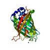
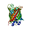
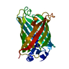
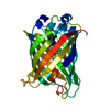
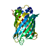
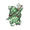
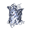
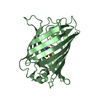
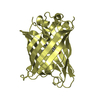
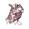
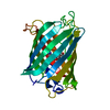
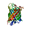
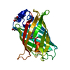
- PDB-4w7e: Crystal Structure of Full-Length Split GFP Mutant E124H/K126H Wit... -
+ Open data

- Basic information
| Entry | Database: PDB / ID: 4w7e | ||||||
|---|---|---|---|---|---|---|---|
| Title | Crystal Structure of Full-Length Split GFP Mutant E124H/K126H With Copper Mediated Crystal Contacts, P 41 21 2 Space Group | ||||||
 Components Components | fluorescent protein D21H/K26H | ||||||
 Keywords Keywords | FLUORESCENT PROTEIN | ||||||
| Function / homology | Green Fluorescent Protein / Green fluorescent protein / Beta Barrel / Mainly Beta / COPPER (II) ION / IMIDAZOLE Function and homology information Function and homology information | ||||||
| Biological species | synthetic construct (others) | ||||||
| Method |  X-RAY DIFFRACTION / SYNCHROTRON / MOLECULAR REPLACEMENT / Resolution: 2.592 Å X-RAY DIFFRACTION / SYNCHROTRON / MOLECULAR REPLACEMENT / Resolution: 2.592 Å | ||||||
 Authors Authors | Leibly, D.J. / Waldo, G.S. / Yeates, T.O. | ||||||
| Funding support |  United States, 1items United States, 1items
| ||||||
 Citation Citation | Journal: Structure / Year: 2015 Title: A Suite of Engineered GFP Molecules for Oligomeric Scaffolding. Authors: Leibly, D.J. / Arbing, M.A. / Pashkov, I. / DeVore, N. / Waldo, G.S. / Terwilliger, T.C. / Yeates, T.O. | ||||||
| History |
|
- Structure visualization
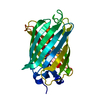
| Structure viewer | Molecule: MolmilJmol/JSmol |
|---|
- Downloads & links
-Download
| PDBx/mmCIF format | 4w7e.cif.gz | 59.5 KB | Display | PDBx/mmCIF format |
|---|---|---|---|---|
| PDB format | pdb4w7e.ent.gz | 42.1 KB | Display | PDB format |
| PDBx/mmJSON format | 4w7e.json.gz | Tree view | PDBx/mmJSON format | |
| Others |  Other downloads Other downloads |
-Validation report
| Summary document | 4w7e_validation.pdf.gz | 440.7 KB | Display | wwPDB validaton report |
|---|---|---|---|---|
| Full document | 4w7e_full_validation.pdf.gz | 443.4 KB | Display | |
| Data in XML | 4w7e_validation.xml.gz | 11.2 KB | Display | |
| Data in CIF | 4w7e_validation.cif.gz | 14.2 KB | Display | |
| Arichive directory | https://data.pdbj.org/pub/pdb/validation_reports/w7/4w7eftp://data.pdbj.org/pub/pdb/validation_reports/w7/4w7e | HTTPS FTP |
-Related structure data
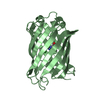
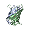
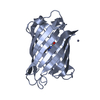
| Related structure data | 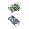 4w69C 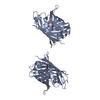 4w6aC 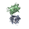 4w6bC 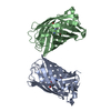 4w6cC 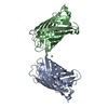 4w6dC 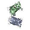 4w6fC 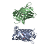 4w6gC 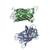 4w6hC  4w6iC  4w6jC  4w6kC 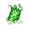 4w6lC  4w6mC  4w6nC  4w6oC  4w6pC  4w6rC  4w6sC 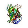 4w6tC 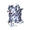 4w6uC 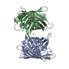 4w72C 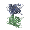 4w73C  4w74C  4w75C 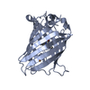 4w76C 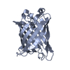 4w77C  4w7aC  4w7cC  4w7dC 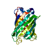 4w7fC  4w7rC  4w7xC  2b3pS C: citing same article ( S: Starting model for refinement |
|---|---|
| Similar structure data |


-Links
 PDBj
PDBj


- Assembly
| Deposited unit | 
| ||||||||
|---|---|---|---|---|---|---|---|---|---|
| 1 |
| ||||||||
| Unit cell |
|
-Components
| #1: Protein | Mass: 26120.402 Da / Num. of mol.: 1 Source method: isolated from a genetically manipulated source Source: (gene. exp.) synthetic construct (others) / Production host:  |
|---|---|
| #2: Chemical | ChemComp-CU /   Mass: 63.546 Da / Num. of mol.: 1 / Source method: obtained synthetically / Formula: Cu Mass: 63.546 Da / Num. of mol.: 1 / Source method: obtained synthetically / Formula: Cu |
| #3: Chemical | ChemComp-IMD /   Mass: 69.085 Da / Num. of mol.: 1 / Source method: obtained synthetically / Formula: C3H5N2 Mass: 69.085 Da / Num. of mol.: 1 / Source method: obtained synthetically / Formula: C3H5N2 |
| #4: Water | ChemComp-HOH /  Mass: 18.015 Da / Num. of mol.: 26 / Source method: isolated from a natural source / Formula: H2O Mass: 18.015 Da / Num. of mol.: 26 / Source method: isolated from a natural source / Formula: H2O |
| Has protein modification | Y |
-Experimental details
-Experiment
| Experiment | Method: X-RAY DIFFRACTION / Number of used crystals: 1 |
|---|
- Sample preparation
| Crystal | Density Matthews: 3.09 Å3/Da / Density % sol: 60.18 % |
|---|---|
| Crystal grow | Temperature: 298 K / Method: vapor diffusion, hanging drop / pH: 8 / Details: 0.1M Imidazole pH 8.0, 10% PEG8000 |
-Data collection
| Diffraction | Mean temperature: 100 K |
|---|---|
| Diffraction source | Source: SYNCHROTRON / Site: APS / Beamline: 24-ID-C / Wavelength: 0.9792 Å |
| Detector | Type: DECTRIS PILATUS 6M-F / Detector: PIXEL / Date: Dec 8, 2013 |
| Radiation | Protocol: SINGLE WAVELENGTH / Monochromatic (M) / Laue (L): M / Scattering type: x-ray |
| Radiation wavelength | Wavelength: 0.9792 Å / Relative weight: 1 |
| Reflection | Resolution: 2.592→67.92 Å / Num. obs: 10580 / % possible obs: 99.7 % / Redundancy: 12.8 % / Rsym value: 0.132 / Net I/σ(I): 19.81 |
- Processing
| Software |
| |||||||||||||||||||||||||||||||||||||||||||||||||||||||||||||||
|---|---|---|---|---|---|---|---|---|---|---|---|---|---|---|---|---|---|---|---|---|---|---|---|---|---|---|---|---|---|---|---|---|---|---|---|---|---|---|---|---|---|---|---|---|---|---|---|---|---|---|---|---|---|---|---|---|---|---|---|---|---|---|---|---|
| Refinement | Method to determine structure: MOLECULAR REPLACEMENT Starting model: 2B3P Resolution: 2.592→67.92 Å / SU ML: 0.42 / Cross valid method: FREE R-VALUE / σ(F): 1.36 / Phase error: 33.5 / Stereochemistry target values: ML
| |||||||||||||||||||||||||||||||||||||||||||||||||||||||||||||||
| Solvent computation | Shrinkage radii: 0.9 Å / VDW probe radii: 1.11 Å / Solvent model: FLAT BULK SOLVENT MODEL | |||||||||||||||||||||||||||||||||||||||||||||||||||||||||||||||
| Refinement step | Cycle: LAST / Resolution: 2.592→67.92 Å
| |||||||||||||||||||||||||||||||||||||||||||||||||||||||||||||||
| Refine LS restraints |
| |||||||||||||||||||||||||||||||||||||||||||||||||||||||||||||||
| LS refinement shell |
|
