 Movie
Movie Controller


+ Open data

- Basic information
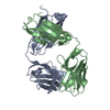
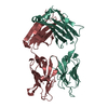
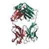
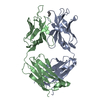
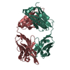
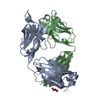
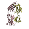
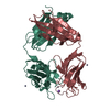
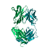
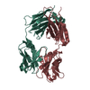
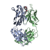
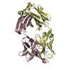
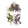
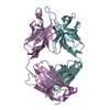
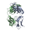
| Entry | Database: PDB / ID: 4uin | ||||||
|---|---|---|---|---|---|---|---|
| Title | crystal structure of quinine-dependent Fab 314.3 with quinine | ||||||
 Components Components | (FAB 314.3) x 2 | ||||||
 Keywords Keywords | IMMUNE SYSTEM / QUININE-DEPENDENT / MOUSE MAB | ||||||
| Function / homology | Immunoglobulins / Immunoglobulin-like / Sandwich / Mainly Beta / Quinine Function and homology information Function and homology information | ||||||
| Biological species |  | ||||||
| Method |  X-RAY DIFFRACTION / SYNCHROTRON / MOLECULAR REPLACEMENT / Resolution: 2.5 Å X-RAY DIFFRACTION / SYNCHROTRON / MOLECULAR REPLACEMENT / Resolution: 2.5 Å | ||||||
 Authors Authors | Zhu, J. / Zhu, J. / Bougie, D.W. / Aster, R.H. / Springer, T.A. | ||||||
 Citation Citation | Journal: Blood / Year: 2015 Title: Structural Basis for Quinine-Dependent Antibody Binding to Platelet Integrin Alphaiib Beta3 Authors: Zhu, J. / Zhu, J. / Bougie, D.W. / Aster, R.H. / Springer, T.A. | ||||||
| History |
|
- Structure visualization
| Structure viewer | Molecule: MolmilJmol/JSmol |
|---|
- Downloads & links
-Download
| PDBx/mmCIF format | 4uin.cif.gz | 250 KB | Display | PDBx/mmCIF format |
|---|---|---|---|---|
| PDB format | pdb4uin.ent.gz | 206.6 KB | Display | PDB format |
| PDBx/mmJSON format | 4uin.json.gz | Tree view | PDBx/mmJSON format | |
| Others |  Other downloads Other downloads |
-Validation report
| Arichive directory | https://data.pdbj.org/pub/pdb/validation_reports/ui/4uinftp://data.pdbj.org/pub/pdb/validation_reports/ui/4uin | HTTPS FTP |
|---|
-Related structure data
| Related structure data |  4uikSC 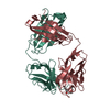 4uilC  4uimC S: Starting model for refinement C: citing same article ( |
|---|---|
| Similar structure data |


-Links
 PDBj
PDBj

- Assembly
| Deposited unit | 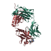
| ||||||||
|---|---|---|---|---|---|---|---|---|---|
| 1 |
| ||||||||
| Unit cell |
|
-Components
| #1: Antibody | Mass: 24257.135 Da / Num. of mol.: 1 / Fragment: HEAVY CHAIN, RESIDUES 1-225 / Source method: isolated from a natural source / Details: ASCITES FLUID / Source: (natural) |
|---|---|
| #2: Antibody | Mass: 23590.900 Da / Num. of mol.: 1 / Fragment: LIGHT CHAIN, RESIDUES 1-214 / Source method: isolated from a natural source / Details: ASCITES FLUID / Source: (natural) |
| #3: Chemical | ChemComp-QI9 / 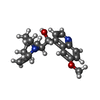  Mass: 324.417 Da / Num. of mol.: 1 / Source method: obtained synthetically / Formula: C20H24N2O2 Mass: 324.417 Da / Num. of mol.: 1 / Source method: obtained synthetically / Formula: C20H24N2O2 |
| #4: Water | ChemComp-HOH /  Mass: 18.015 Da / Num. of mol.: 90 / Source method: isolated from a natural source / Formula: H2O Mass: 18.015 Da / Num. of mol.: 90 / Source method: isolated from a natural source / Formula: H2O |
| Has protein modification | Y |
-Experimental details
-Experiment
| Experiment | Method: X-RAY DIFFRACTION / Number of used crystals: 1 |
|---|
- Sample preparation
| Crystal | Density Matthews: 2.24 Å3/Da / Density % sol: 45.04 % / Description: NONE |
|---|---|
| Crystal grow | pH: 7.5 / Details: 25% PEG 1500, PH 7.5 |
-Data collection
| Diffraction | Mean temperature: 100 K |
|---|---|
| Diffraction source | Source: SYNCHROTRON / Site: APS  / Beamline: 23-ID-D / Wavelength: 1.03321 / Beamline: 23-ID-D / Wavelength: 1.03321 |
| Detector | Type: MARRESEARCH / Detector: CCD / Date: Apr 10, 2012 / Details: DOUBLE MIRROR |
| Radiation | Monochromator: SI CRYSTAL / Protocol: SINGLE WAVELENGTH / Monochromatic (M) / Laue (L): M / Scattering type: x-ray |
| Radiation wavelength | Wavelength: 1.03321 Å / Relative weight: 1 |
| Reflection | Resolution: 2.5→50 Å / Num. obs: 14747 / % possible obs: 99.3 % / Observed criterion σ(I): 0 / Redundancy: 3.7 % / Biso Wilson estimate: 26.11 Å2 / Rmerge(I) obs: 0.13 / Net I/σ(I): 9.6 |
| Reflection shell | Resolution: 2.5→2.59 Å / Redundancy: 3 % / Rmerge(I) obs: 0.76 / Mean I/σ(I) obs: 1.5 / % possible all: 95.6 |
- Processing
| Software |
| ||||||||||||||||||||||||||||||||||||||||||||||||||||||||||||||||||||||||||||||||||||||||||||||||||||||||||||||||||||||||||||||||||||||||||||||||||||||
|---|---|---|---|---|---|---|---|---|---|---|---|---|---|---|---|---|---|---|---|---|---|---|---|---|---|---|---|---|---|---|---|---|---|---|---|---|---|---|---|---|---|---|---|---|---|---|---|---|---|---|---|---|---|---|---|---|---|---|---|---|---|---|---|---|---|---|---|---|---|---|---|---|---|---|---|---|---|---|---|---|---|---|---|---|---|---|---|---|---|---|---|---|---|---|---|---|---|---|---|---|---|---|---|---|---|---|---|---|---|---|---|---|---|---|---|---|---|---|---|---|---|---|---|---|---|---|---|---|---|---|---|---|---|---|---|---|---|---|---|---|---|---|---|---|---|---|---|---|---|---|---|
| Refinement | Method to determine structure: MOLECULAR REPLACEMENT Starting model: PDB ENTRY 4UIK Resolution: 2.5→43.771 Å / SU ML: 0.32 / σ(F): 1.99 / Phase error: 25.39 / Stereochemistry target values: ML
| ||||||||||||||||||||||||||||||||||||||||||||||||||||||||||||||||||||||||||||||||||||||||||||||||||||||||||||||||||||||||||||||||||||||||||||||||||||||
| Solvent computation | Shrinkage radii: 0.7 Å / VDW probe radii: 1 Å / Solvent model: FLAT BULK SOLVENT MODEL | ||||||||||||||||||||||||||||||||||||||||||||||||||||||||||||||||||||||||||||||||||||||||||||||||||||||||||||||||||||||||||||||||||||||||||||||||||||||
| Displacement parameters | Biso mean: 30 Å2 | ||||||||||||||||||||||||||||||||||||||||||||||||||||||||||||||||||||||||||||||||||||||||||||||||||||||||||||||||||||||||||||||||||||||||||||||||||||||
| Refinement step | Cycle: LAST / Resolution: 2.5→43.771 Å
| ||||||||||||||||||||||||||||||||||||||||||||||||||||||||||||||||||||||||||||||||||||||||||||||||||||||||||||||||||||||||||||||||||||||||||||||||||||||
| Refine LS restraints |
| ||||||||||||||||||||||||||||||||||||||||||||||||||||||||||||||||||||||||||||||||||||||||||||||||||||||||||||||||||||||||||||||||||||||||||||||||||||||
| LS refinement shell |
| ||||||||||||||||||||||||||||||||||||||||||||||||||||||||||||||||||||||||||||||||||||||||||||||||||||||||||||||||||||||||||||||||||||||||||||||||||||||
| Refinement TLS params. | Method: refined / Refine-ID: X-RAY DIFFRACTION
| ||||||||||||||||||||||||||||||||||||||||||||||||||||||||||||||||||||||||||||||||||||||||||||||||||||||||||||||||||||||||||||||||||||||||||||||||||||||
| Refinement TLS group |
|
