 Movie
Movie Controller

[English] 日本語
 Yorodumi
- PDB-4u0y: Crystal structure of the DNA-binding domains of YvoA in complex w... -
+ Open data

- Basic information
| Entry | Database: PDB / ID: 4u0y | |||||||||
|---|---|---|---|---|---|---|---|---|---|---|
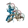
| Title | Crystal structure of the DNA-binding domains of YvoA in complex with palindromic operator DNA | |||||||||
 Components Components |
| |||||||||
 Keywords Keywords | TRANSCRIPTION / Repressor / Bacterial transcription regulation / Transcription factor / GntR/HutC family / Winged helix-turn-helix motif / N-acetylglucosamine utilization / DNA-binding / Operator-binding | |||||||||
| Function / homology |  Function and homology information Function and homology informationDNA-binding transcription factor activity / negative regulation of DNA-templated transcription / DNA binding Similarity search - Function | |||||||||
| Biological species |  | |||||||||
| Method |  X-RAY DIFFRACTION / SYNCHROTRON / MOLECULAR REPLACEMENT / Resolution: 1.91 Å X-RAY DIFFRACTION / SYNCHROTRON / MOLECULAR REPLACEMENT / Resolution: 1.91 Å | |||||||||
 Authors Authors | Fillenberg, S.B. / Muller, Y.A. | |||||||||
| Funding support |  Germany, 1items Germany, 1items
| |||||||||
 Citation Citation | Journal: Nucleic Acids Res. / Year: 2015 Title: Structural insight into operator dre-sites recognition and effector binding in the GntR/HutC transcription regulator NagR. Authors: Fillenberg, S.B. / Grau, F.C. / Seidel, G. / Muller, Y.A. | |||||||||
| History |
|
- Structure visualization
| Structure viewer | Molecule: MolmilJmol/JSmol |
|---|
- Downloads & links
-Download
| PDBx/mmCIF format | 4u0y.cif.gz | 98.8 KB | Display | PDBx/mmCIF format |
|---|---|---|---|---|
| PDB format | pdb4u0y.ent.gz | 71.3 KB | Display | PDB format |
| PDBx/mmJSON format | 4u0y.json.gz | Tree view | PDBx/mmJSON format | |
| Others |  Other downloads Other downloads |
-Validation report
| Arichive directory | https://data.pdbj.org/pub/pdb/validation_reports/u0/4u0yftp://data.pdbj.org/pub/pdb/validation_reports/u0/4u0y | HTTPS FTP |
|---|
-Related structure data
| Related structure data |  4u0vC  4u0wC  4wwcC 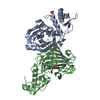 2wv0S S: Starting model for refinement C: citing same article ( |
|---|---|
| Similar structure data |


-Links
 PDBj
PDBj


- Assembly
| Deposited unit | 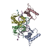
| ||||||||
|---|---|---|---|---|---|---|---|---|---|
| 1 |
| ||||||||
| Unit cell |
|
-Components
| #1: Protein | Mass: 8965.283 Da / Num. of mol.: 4 / Fragment: UNP residues 1-75 Source method: isolated from a genetically manipulated source Details: Truncated form of YvoA; comprises only the DNA-binding domain (residues 1-75) Source: (gene. exp.) Gene: yvoA, BSU35030 / Production host: #2: DNA chain | Mass: 4584.984 Da / Num. of mol.: 2 / Source method: obtained synthetically Details: 15mer palindromic dsDNA construct; derived from the consensus sequence of the two native non-palindromic dre-site sequences upstream of nagAB-yvoA and nagP Source: (synth.) #3: Chemical |   Mass: 35.453 Da / Num. of mol.: 2 / Source method: obtained synthetically / Formula: Cl Mass: 35.453 Da / Num. of mol.: 2 / Source method: obtained synthetically / Formula: Cl#4: Water | ChemComp-HOH / |  Mass: 18.015 Da / Num. of mol.: 287 / Source method: isolated from a natural source / Formula: H2O Mass: 18.015 Da / Num. of mol.: 287 / Source method: isolated from a natural source / Formula: H2O |
|---|
-Experimental details
-Experiment
| Experiment | Method: X-RAY DIFFRACTION / Number of used crystals: 1 |
|---|
- Sample preparation
| Crystal | Density Matthews: 2.39 Å3/Da / Density % sol: 48.46 % |
|---|---|
| Crystal grow | Temperature: 292.15 K / Method: vapor diffusion, sitting drop / pH: 6.5 Details: 50 mM sodium cacodylate, 200 mM sodium citrate, 10 mM MgCl2, 5 % (v/v) isopropanol |
-Data collection
| Diffraction | Mean temperature: 100 K |
|---|---|
| Diffraction source | Source: SYNCHROTRON / Site: BESSY / Beamline: 14.1 / Wavelength: 0.91841 Å |
| Detector | Type: RAYONIX MX-225 / Detector: CCD / Date: Oct 8, 2010 |
| Radiation | Monochromator: Si-111 crystal / Protocol: SINGLE WAVELENGTH / Monochromatic (M) / Laue (L): M / Scattering type: x-ray |
| Radiation wavelength | Wavelength: 0.91841 Å / Relative weight: 1 |
| Reflection | Resolution: 1.91→32.52 Å / Num. obs: 31230 / % possible obs: 99.8 % / Redundancy: 3.8 % / Biso Wilson estimate: 25.86 Å2 / Rmerge(I) obs: 0.085 / Net I/σ(I): 10.61 |
| Reflection shell | Resolution: 1.91→1.98 Å / Redundancy: 3.7 % / Rmerge(I) obs: 0.87 / Mean I/σ(I) obs: 1.9 / % possible all: 98.7 |
- Processing
| Software |
| ||||||||||||||||||||||||||||||||||||||||||||||||||||||||||||||||||||||||||||||||||||
|---|---|---|---|---|---|---|---|---|---|---|---|---|---|---|---|---|---|---|---|---|---|---|---|---|---|---|---|---|---|---|---|---|---|---|---|---|---|---|---|---|---|---|---|---|---|---|---|---|---|---|---|---|---|---|---|---|---|---|---|---|---|---|---|---|---|---|---|---|---|---|---|---|---|---|---|---|---|---|---|---|---|---|---|---|---|
| Refinement | Method to determine structure: MOLECULAR REPLACEMENT Starting model: DNA-binding domain of PDB entry 2wv0 Resolution: 1.91→32.52 Å / Occupancy max: 1 / Occupancy min: 0.45 / FOM work R set: 0.8245 / SU ML: 0.23 / Cross valid method: FREE R-VALUE / σ(F): 1.95 / Phase error: 25.14 / Stereochemistry target values: ML
| ||||||||||||||||||||||||||||||||||||||||||||||||||||||||||||||||||||||||||||||||||||
| Solvent computation | Shrinkage radii: 0.9 Å / VDW probe radii: 1.11 Å / Solvent model: FLAT BULK SOLVENT MODEL | ||||||||||||||||||||||||||||||||||||||||||||||||||||||||||||||||||||||||||||||||||||
| Displacement parameters | Biso max: 99.38 Å2 / Biso mean: 29.8537 Å2 / Biso min: 13.23 Å2 | ||||||||||||||||||||||||||||||||||||||||||||||||||||||||||||||||||||||||||||||||||||
| Refinement step | Cycle: LAST / Resolution: 1.91→32.52 Å
| ||||||||||||||||||||||||||||||||||||||||||||||||||||||||||||||||||||||||||||||||||||
| Refine LS restraints |
| ||||||||||||||||||||||||||||||||||||||||||||||||||||||||||||||||||||||||||||||||||||
| LS refinement shell | Refine-ID: X-RAY DIFFRACTION / Total num. of bins used: 11
|
