 Movie
Movie Controller

[English] 日本語
 Yorodumi
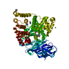
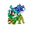
- PDB-6m7t: Human DNA polymerase eta in a non-productive ternary complex with... -
+ Open data

- Basic information
| Entry | Database: PDB / ID: 6m7t | ||||||
|---|---|---|---|---|---|---|---|
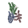
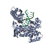
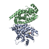
| Title | Human DNA polymerase eta in a non-productive ternary complex with Ca2+ and dTTP oppositing cdA | ||||||
 Components Components |
| ||||||
 Keywords Keywords | TRANSFERASE/DNA / TRANSLESION SYNTHESIS / cdA / DNA BINDING PROTEIN / TRANSFERASE-DNA complex | ||||||
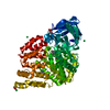
| Function / homology |  Function and homology information Function and homology informationresponse to UV-C / error-free translesion synthesis / DNA synthesis involved in DNA repair / cellular response to UV-C / pyrimidine dimer repair / error-prone translesion synthesis / regulation of DNA repair / replication fork / Termination of translesion DNA synthesis / response to radiation ...response to UV-C / error-free translesion synthesis / DNA synthesis involved in DNA repair / cellular response to UV-C / pyrimidine dimer repair / error-prone translesion synthesis / regulation of DNA repair / replication fork / Termination of translesion DNA synthesis / response to radiation / Translesion Synthesis by POLH / HDR through Homologous Recombination (HRR) / site of double-strand break / DNA-directed DNA polymerase / damaged DNA binding / DNA-directed DNA polymerase activity / DNA replication / DNA repair / zinc ion binding / nucleoplasm / nucleus / cytosol Similarity search - Function | ||||||
| Biological species |  Homo sapiens (human) Homo sapiens (human) | ||||||
| Method |  X-RAY DIFFRACTION / SYNCHROTRON / MOLECULAR REPLACEMENT / Resolution: 2.8 Å X-RAY DIFFRACTION / SYNCHROTRON / MOLECULAR REPLACEMENT / Resolution: 2.8 Å | ||||||
 Authors Authors | Weng, P. / Gao, Y. / Yang, W. | ||||||
| Funding support |  United States, 1items United States, 1items
| ||||||
 Citation Citation | Journal: Proc. Natl. Acad. Sci. U.S.A. / Year: 2018 Title: Bypassing a 8,5'-cyclo-2'-deoxyadenosine lesion by human DNA polymerase eta at atomic resolution. Authors: Weng, P.J. / Gao, Y. / Gregory, M.T. / Wang, P. / Wang, Y. / Yang, W. | ||||||
| History |
|
- Structure visualization
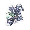
| Structure viewer | Molecule: MolmilJmol/JSmol |
|---|
- Downloads & links
-Download
| PDBx/mmCIF format | 6m7t.cif.gz | 116.5 KB | Display | PDBx/mmCIF format |
|---|---|---|---|---|
| PDB format | pdb6m7t.ent.gz | 83.6 KB | Display | PDB format |
| PDBx/mmJSON format | 6m7t.json.gz | Tree view | PDBx/mmJSON format | |
| Others |  Other downloads Other downloads |
-Validation report
| Arichive directory | https://data.pdbj.org/pub/pdb/validation_reports/m7/6m7tftp://data.pdbj.org/pub/pdb/validation_reports/m7/6m7t | HTTPS FTP |
|---|
-Related structure data
| Related structure data |  6m7oC  6m7pC  6m7uC  6m7vC  3si8S S: Starting model for refinement C: citing same article ( |
|---|---|
| Similar structure data |


-Links
 PDBj
PDBj


- Assembly
| Deposited unit | 
| ||||||||
|---|---|---|---|---|---|---|---|---|---|
| 1 |
| ||||||||
| Unit cell |
|
-Components
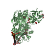
-Protein , 1 types, 1 molecules A
| #1: Protein | Mass: 48617.707 Da / Num. of mol.: 1 / Fragment: residues 1-432 Source method: isolated from a genetically manipulated source Source: (gene. exp.) Homo sapiens (human) / Gene: POLH, RAD30, RAD30A, XPV / Production host:  |
|---|
-DNA chain , 2 types, 2 molecules PT
| #2: DNA chain | Mass: 2506.665 Da / Num. of mol.: 1 / Source method: obtained synthetically / Source: (synth.) Homo sapiens (human) |
|---|---|
| #3: DNA chain | Mass: 3275.159 Da / Num. of mol.: 1 / Source method: obtained synthetically / Source: (synth.) Homo sapiens (human) |
-Non-polymers , 6 types, 40 molecules 


| #4: Chemical | ChemComp-NI /  Mass: 58.693 Da / Num. of mol.: 1 / Source method: obtained synthetically / Formula: Ni Mass: 58.693 Da / Num. of mol.: 1 / Source method: obtained synthetically / Formula: Ni | ||
|---|---|---|---|
| #5: Chemical | ChemComp-TTP /  Mass: 482.168 Da / Num. of mol.: 1 / Source method: obtained synthetically / Formula: C10H17N2O14P3 Mass: 482.168 Da / Num. of mol.: 1 / Source method: obtained synthetically / Formula: C10H17N2O14P3 | ||
| #6: Chemical | ChemComp-CA /  Mass: 40.078 Da / Num. of mol.: 1 / Source method: obtained synthetically / Formula: Ca Mass: 40.078 Da / Num. of mol.: 1 / Source method: obtained synthetically / Formula: Ca | ||
| #7: Chemical | ChemComp-EDO /  Mass: 62.068 Da / Num. of mol.: 1 / Source method: isolated from a natural source / Formula: C2H6O2 Mass: 62.068 Da / Num. of mol.: 1 / Source method: isolated from a natural source / Formula: C2H6O2 | ||
| #8: Chemical |  Mass: 92.094 Da / Num. of mol.: 3 / Source method: obtained synthetically / Formula: C3H8O3 Mass: 92.094 Da / Num. of mol.: 3 / Source method: obtained synthetically / Formula: C3H8O3#9: Water | ChemComp-HOH / | Mass: 18.015 Da / Num. of mol.: 33 / Source method: isolated from a natural source / Formula: H2O |
-Experimental details
-Experiment
| Experiment | Method: X-RAY DIFFRACTION / Number of used crystals: 1 |
|---|
- Sample preparation
| Crystal | Density Matthews: 3.43 Å3/Da / Density % sol: 64.12 % |
|---|---|
| Crystal grow | Temperature: 295 K / Method: vapor diffusion, hanging drop / Details: 16 % PEG 2000 MME 100 mM MES, pH 6.0 |
-Data collection
| Diffraction | Mean temperature: 100 K |
|---|---|
| Diffraction source | Source: SYNCHROTRON / Site: APS / Beamline: 22-BM / Wavelength: 1 Å |
| Detector | Type: MARMOSAIC 225 mm CCD / Detector: CCD / Date: Aug 14, 2014 |
| Radiation | Protocol: SINGLE WAVELENGTH / Monochromatic (M) / Laue (L): M / Scattering type: x-ray |
| Radiation wavelength | Wavelength: 1 Å / Relative weight: 1 |
| Reflection | Resolution: 2.8→30 Å / Num. obs: 19880 / % possible obs: 99.8 % / Redundancy: 5 % / CC1/2: 0.998 / Rmerge(I) obs: 0.092 / Net I/σ(I): 16 |
| Reflection shell | Resolution: 2.8→2.87 Å / Rmerge(I) obs: 0.643 / Num. unique obs: 1364 / CC1/2: 0.894 |
- Processing
| Software |
| ||||||||||||||||||||||||||||||||||||||||||||||||||||||||||||||||||||||||||||||||||||||||||||||||||
|---|---|---|---|---|---|---|---|---|---|---|---|---|---|---|---|---|---|---|---|---|---|---|---|---|---|---|---|---|---|---|---|---|---|---|---|---|---|---|---|---|---|---|---|---|---|---|---|---|---|---|---|---|---|---|---|---|---|---|---|---|---|---|---|---|---|---|---|---|---|---|---|---|---|---|---|---|---|---|---|---|---|---|---|---|---|---|---|---|---|---|---|---|---|---|---|---|---|---|---|
| Refinement | Method to determine structure: MOLECULAR REPLACEMENT Starting model: 3SI8 Resolution: 2.8→29.734 Å / SU ML: 0.4 / Cross valid method: FREE R-VALUE / σ(F): 0.98 / Phase error: 29.84 / Stereochemistry target values: ML
| ||||||||||||||||||||||||||||||||||||||||||||||||||||||||||||||||||||||||||||||||||||||||||||||||||
| Solvent computation | Shrinkage radii: 0.9 Å / VDW probe radii: 1.11 Å / Solvent model: FLAT BULK SOLVENT MODEL | ||||||||||||||||||||||||||||||||||||||||||||||||||||||||||||||||||||||||||||||||||||||||||||||||||
| Refinement step | Cycle: LAST / Resolution: 2.8→29.734 Å
| ||||||||||||||||||||||||||||||||||||||||||||||||||||||||||||||||||||||||||||||||||||||||||||||||||
| Refine LS restraints |
| ||||||||||||||||||||||||||||||||||||||||||||||||||||||||||||||||||||||||||||||||||||||||||||||||||
| LS refinement shell |
|
