 Movie
Movie Controller

[English] 日本語
 Yorodumi
- PDB-4pxh: Structure of P450sky (CYP163B3), a cytochrome P450 from skyllamyc... -
+ Open data

- Basic information
| Entry | Database: PDB / ID: 4pxh | ||||||
|---|---|---|---|---|---|---|---|
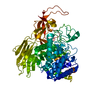
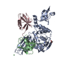
| Title | Structure of P450sky (CYP163B3), a cytochrome P450 from skyllamycin biosynthesis in complex with a peptidyl carrier protein domain | ||||||
 Components Components |
| ||||||
 Keywords Keywords | OXIDOREDUCTASE/PROTEIN BINDING / Cytochrome P450 fold / beta-aminoacyl carrier protein hydroxylase / peptidyl carrier protein domains / skyllamycin NRPS / OXIDOREDUCTASE-PROTEIN BINDING complex | ||||||
| Function / homology |  Function and homology information Function and homology informationmonocarboxylic acid biosynthetic process / cholest-4-en-3-one 26-monooxygenase activity / amino acid activation for nonribosomal peptide biosynthetic process / steroid hydroxylase activity / cholesterol catabolic process / secondary metabolite biosynthetic process / lipid biosynthetic process / catalytic activity / phosphopantetheine binding / antibiotic biosynthetic process ...monocarboxylic acid biosynthetic process / cholest-4-en-3-one 26-monooxygenase activity / amino acid activation for nonribosomal peptide biosynthetic process / steroid hydroxylase activity / cholesterol catabolic process / secondary metabolite biosynthetic process / lipid biosynthetic process / catalytic activity / phosphopantetheine binding / antibiotic biosynthetic process / iron ion binding / heme binding / cytosol Similarity search - Function | ||||||
| Biological species |  Streptomyces sp. Acta 2897 (bacteria) Streptomyces sp. Acta 2897 (bacteria) | ||||||
| Method |  X-RAY DIFFRACTION / SYNCHROTRON / MOLECULAR REPLACEMENT / Resolution: 2.7 Å X-RAY DIFFRACTION / SYNCHROTRON / MOLECULAR REPLACEMENT / Resolution: 2.7 Å | ||||||
 Authors Authors | Haslinger, K. / Cryle, M.J. | ||||||
 Citation Citation | Journal: Angew.Chem.Int.Ed.Engl. / Year: 2014 Title: The structure of a transient complex of a nonribosomal Peptide synthetase and a cytochrome p450 monooxygenase. Authors: Haslinger, K. / Brieke, C. / Uhlmann, S. / Sieverling, L. / Sussmuth, R.D. / Cryle, M.J. | ||||||
| History |
|
- Structure visualization
| Structure viewer | Molecule: MolmilJmol/JSmol |
|---|
- Downloads & links
-Download
| PDBx/mmCIF format | 4pxh.cif.gz | 586.5 KB | Display | PDBx/mmCIF format |
|---|---|---|---|---|
| PDB format | pdb4pxh.ent.gz | 487.8 KB | Display | PDB format |
| PDBx/mmJSON format | 4pxh.json.gz | Tree view | PDBx/mmJSON format | |
| Others |  Other downloads Other downloads |
-Validation report
| Arichive directory | https://data.pdbj.org/pub/pdb/validation_reports/px/4pxhftp://data.pdbj.org/pub/pdb/validation_reports/px/4pxh | HTTPS FTP |
|---|
-Related structure data
| Related structure data | 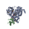 4pwvSC S: Starting model for refinement C: citing same article ( |
|---|---|
| Similar structure data |


-Links
 PDBj
PDBj


- Assembly
| Deposited unit | 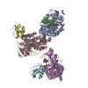
| ||||||||
|---|---|---|---|---|---|---|---|---|---|
| 1 | 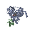
| ||||||||
| 2 | 
| ||||||||
| 3 | 
| ||||||||
| Unit cell |
|
-Components
| #1: Protein | Mass: 49369.414 Da / Num. of mol.: 3 Source method: isolated from a genetically manipulated source Source: (gene. exp.) Streptomyces sp. Acta 2897 (bacteria) / Strain: Acta 2897 / Gene: sky32 / Plasmid: pET28a(+) / Production host: #2: Protein | Mass: 10186.228 Da / Num. of mol.: 3 / Fragment: peptidyl carrier protein domain Source method: isolated from a genetically manipulated source Source: (gene. exp.) Streptomyces sp. Acta 2897 (bacteria) / Strain: Acta 2897 / Gene: sky30 / Plasmid: pET28a(+) / Production host: #3: Chemical |   Mass: 616.487 Da / Num. of mol.: 3 / Source method: obtained synthetically / Formula: C34H32FeN4O4 Mass: 616.487 Da / Num. of mol.: 3 / Source method: obtained synthetically / Formula: C34H32FeN4O4#4: Chemical |   Mass: 452.420 Da / Num. of mol.: 3 / Source method: obtained synthetically / Formula: C15H25N4O8PS Mass: 452.420 Da / Num. of mol.: 3 / Source method: obtained synthetically / Formula: C15H25N4O8PS#5: Water | ChemComp-HOH / |  Mass: 18.015 Da / Num. of mol.: 73 / Source method: isolated from a natural source / Formula: H2O Mass: 18.015 Da / Num. of mol.: 73 / Source method: isolated from a natural source / Formula: H2OHas protein modification | Y | |
|---|
-Experimental details
-Experiment
| Experiment | Method: X-RAY DIFFRACTION / Number of used crystals: 1 |
|---|
- Sample preparation
| Crystal | Density Matthews: 3.56 Å3/Da / Density % sol: 65.49 % |
|---|---|
| Crystal grow | Temperature: 293 K / Method: vapor diffusion, hanging drop / pH: 8.5 Details: 20% PEG3350, 0.15 M calcium acetate, 0.1 M Tris, pH 8.5, VAPOR DIFFUSION, HANGING DROP, temperature 293K |
-Data collection
| Diffraction | Mean temperature: 100 K |
|---|---|
| Diffraction source | Source: SYNCHROTRON / Site: SLS  / Beamline: X10SA / Wavelength: 1.00002 Å / Beamline: X10SA / Wavelength: 1.00002 Å |
| Detector | Type: DECTRIS PILATUS 6M / Detector: PIXEL / Date: Jul 18, 2013 |
| Radiation | Monochromator: double crystal Si(111) / Protocol: SINGLE WAVELENGTH / Monochromatic (M) / Laue (L): M / Scattering type: x-ray |
| Radiation wavelength | Wavelength: 1.00002 Å / Relative weight: 1 |
| Reflection | Resolution: 2.7→47.893 Å / Num. all: 72148 / Num. obs: 72098 / % possible obs: 99.9 % / Observed criterion σ(I): 3 / Redundancy: 19.7 % / Rmerge(I) obs: 0.117 / Net I/σ(I): 24.5 |
| Reflection shell | Resolution: 2.7→3 Å / Redundancy: 19.9 % / Rmerge(I) obs: 0.661 / Mean I/σ(I) obs: 5.6 / % possible all: 100 |
- Processing
| Software |
| ||||||||||||||||||||||||||||||||||||||||||||||||||||||||||||||||||||||||||||||||||||||||||||||||||||||||||||||||||||||||||||||||||||||||||||||||||||||||||||||||||||||||||||||||||||||
|---|---|---|---|---|---|---|---|---|---|---|---|---|---|---|---|---|---|---|---|---|---|---|---|---|---|---|---|---|---|---|---|---|---|---|---|---|---|---|---|---|---|---|---|---|---|---|---|---|---|---|---|---|---|---|---|---|---|---|---|---|---|---|---|---|---|---|---|---|---|---|---|---|---|---|---|---|---|---|---|---|---|---|---|---|---|---|---|---|---|---|---|---|---|---|---|---|---|---|---|---|---|---|---|---|---|---|---|---|---|---|---|---|---|---|---|---|---|---|---|---|---|---|---|---|---|---|---|---|---|---|---|---|---|---|---|---|---|---|---|---|---|---|---|---|---|---|---|---|---|---|---|---|---|---|---|---|---|---|---|---|---|---|---|---|---|---|---|---|---|---|---|---|---|---|---|---|---|---|---|---|---|---|---|
| Refinement | Method to determine structure: MOLECULAR REPLACEMENT Starting model: PDB ENTRY 4PWV Resolution: 2.7→47.893 Å / Cor.coef. Fo:Fc: 0.933 / Cor.coef. Fo:Fc free: 0.913 / SU B: 19.736 / SU ML: 0.201 / Cross valid method: THROUGHOUT / ESU R: 0.435 / ESU R Free: 0.28 / Stereochemistry target values: MAXIMUM LIKELIHOOD / Details: HYDROGENS HAVE BEEN ADDED IN THE RIDING POSITIONS
| ||||||||||||||||||||||||||||||||||||||||||||||||||||||||||||||||||||||||||||||||||||||||||||||||||||||||||||||||||||||||||||||||||||||||||||||||||||||||||||||||||||||||||||||||||||||
| Solvent computation | Ion probe radii: 0.8 Å / Shrinkage radii: 0.8 Å / VDW probe radii: 1.4 Å / Solvent model: MASK | ||||||||||||||||||||||||||||||||||||||||||||||||||||||||||||||||||||||||||||||||||||||||||||||||||||||||||||||||||||||||||||||||||||||||||||||||||||||||||||||||||||||||||||||||||||||
| Displacement parameters | Biso mean: 54.081 Å2
| ||||||||||||||||||||||||||||||||||||||||||||||||||||||||||||||||||||||||||||||||||||||||||||||||||||||||||||||||||||||||||||||||||||||||||||||||||||||||||||||||||||||||||||||||||||||
| Refinement step | Cycle: LAST / Resolution: 2.7→47.893 Å
| ||||||||||||||||||||||||||||||||||||||||||||||||||||||||||||||||||||||||||||||||||||||||||||||||||||||||||||||||||||||||||||||||||||||||||||||||||||||||||||||||||||||||||||||||||||||
| Refine LS restraints |
|