 Movie
Movie Controller


+ Open data

- Basic information
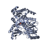
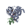
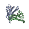
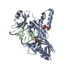
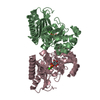
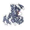
| Entry | Database: PDB / ID: 4eql | ||||||
|---|---|---|---|---|---|---|---|
| Title | Crystal Structure of GH3.12 in complex with AMP and salicylate | ||||||
 Components Components | 4-substituted benzoates-glutamate ligase GH3.12 | ||||||
 Keywords Keywords | LIGASE / firefly luciferase family / acyl adenylase / amino acid conjugation | ||||||
| Function / homology |  Function and homology information Function and homology informationN-(4-aminobenzoyl)-L-glutamate synthetase activity / N-benzoyl-L-glutamate synthetase activity / N-vanillate-L-glutamate synthetase activity / N-(4-hydroxybenzoyl)-L-glutamate synthetase activity / positive regulation of plant-type hypersensitive response / benzoate metabolic process / regulation of systemic acquired resistance / salicylic acid mediated signaling pathway / detection of fungus / acid-amino acid ligase activity ...N-(4-aminobenzoyl)-L-glutamate synthetase activity / N-benzoyl-L-glutamate synthetase activity / N-vanillate-L-glutamate synthetase activity / N-(4-hydroxybenzoyl)-L-glutamate synthetase activity / positive regulation of plant-type hypersensitive response / benzoate metabolic process / regulation of systemic acquired resistance / salicylic acid mediated signaling pathway / detection of fungus / acid-amino acid ligase activity / Ligases; Forming carbon-nitrogen bonds; Acid-amino-acid ligases (peptide synthases) / plant-type hypersensitive response / defense response / cellular response to hypoxia / defense response to bacterium / cytoplasm Similarity search - Function | ||||||
| Biological species |  | ||||||
| Method |  X-RAY DIFFRACTION / SYNCHROTRON / MOLECULAR REPLACEMENT / Resolution: 1.8 Å X-RAY DIFFRACTION / SYNCHROTRON / MOLECULAR REPLACEMENT / Resolution: 1.8 Å | ||||||
 Authors Authors | Westfall, C. / Zubieta, C. / Nanao, M. / Herrmann, J. / Jez, J. | ||||||
 Citation Citation | Journal: Science / Year: 2012 Title: Structural basis for prereceptor modulation of plant hormones by GH3 proteins. Authors: Westfall, C.S. / Zubieta, C. / Herrmann, J. / Kapp, U. / Nanao, M.H. / Jez, J.M. | ||||||
| History |
|
- Structure visualization
| Structure viewer | Molecule: MolmilJmol/JSmol |
|---|
- Downloads & links
-Download
| PDBx/mmCIF format | 4eql.cif.gz | 423.8 KB | Display | PDBx/mmCIF format |
|---|---|---|---|---|
| PDB format | pdb4eql.ent.gz | 341.9 KB | Display | PDB format |
| PDBx/mmJSON format | 4eql.json.gz | Tree view | PDBx/mmJSON format | |
| Others |  Other downloads Other downloads |
-Validation report
| Arichive directory | https://data.pdbj.org/pub/pdb/validation_reports/eq/4eqlftp://data.pdbj.org/pub/pdb/validation_reports/eq/4eql | HTTPS FTP |
|---|
-Related structure data
| Related structure data |  4eplC  4epmC  4eq4SC  4ewvC S: Starting model for refinement C: citing same article ( |
|---|---|
| Similar structure data |


-Links
 PDBj
PDBj


- Assembly
| Deposited unit | 
| ||||||||
|---|---|---|---|---|---|---|---|---|---|
| 1 | 
| ||||||||
| 2 | 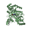
| ||||||||
| 3 |
| ||||||||
| Unit cell |
|
-Components
| #1: Protein | Mass: 65762.430 Da / Num. of mol.: 2 Source method: isolated from a genetically manipulated source Source: (gene. exp.) Gene: GH3.12, GDG1, PBS3, WIN3, At5g13320, T22N19.5, T31B5.140 Production host:  References: UniProt: Q9LYU4, Ligases; Forming carbon-nitrogen bonds; Acid-amino-acid ligases (peptide synthases) #2: Chemical |   Mass: 347.221 Da / Num. of mol.: 2 / Source method: obtained synthetically / Formula: C10H14N5O7P / Comment: AMP*YM Mass: 347.221 Da / Num. of mol.: 2 / Source method: obtained synthetically / Formula: C10H14N5O7P / Comment: AMP*YM#3: Chemical | 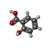  Mass: 138.121 Da / Num. of mol.: 2 / Source method: obtained synthetically / Formula: C7H6O3 Mass: 138.121 Da / Num. of mol.: 2 / Source method: obtained synthetically / Formula: C7H6O3#4: Water | ChemComp-HOH / |  Mass: 18.015 Da / Num. of mol.: 1171 / Source method: isolated from a natural source / Formula: H2O Mass: 18.015 Da / Num. of mol.: 1171 / Source method: isolated from a natural source / Formula: H2O |
|---|
-Experimental details
-Experiment
| Experiment | Method: X-RAY DIFFRACTION / Number of used crystals: 1 |
|---|
- Sample preparation
| Crystal | Density Matthews: 2.26 Å3/Da / Density % sol: 45.59 % |
|---|---|
| Crystal grow | Temperature: 298 K / Method: vapor diffusion, hanging drop / pH: 4.5 Details: 20% PEG3350, 0.25M ammonium acetate, pH 4.5, VAPOR DIFFUSION, HANGING DROP, temperature 298K |
-Data collection
| Diffraction | Mean temperature: 100 K |
|---|---|
| Diffraction source | Source: SYNCHROTRON / Site: ESRF  / Beamline: ID23-2 / Wavelength: 0.873 Å / Beamline: ID23-2 / Wavelength: 0.873 Å |
| Detector | Type: MARMOSAIC 225 mm CCD / Detector: CCD / Date: Feb 23, 2011 |
| Radiation | Monochromator: horizontally side diffracting Silicon 111 crystal Protocol: SINGLE WAVELENGTH / Monochromatic (M) / Laue (L): M / Scattering type: x-ray |
| Radiation wavelength | Wavelength: 0.873 Å / Relative weight: 1 |
| Reflection | Resolution: 1.8→96 Å / Num. all: 108026 / Num. obs: 106773 / % possible obs: 98.8 % / Observed criterion σ(F): 1.9 / Observed criterion σ(I): 1.9 / Redundancy: 3.7 % / Biso Wilson estimate: 25.6 Å2 / Rmerge(I) obs: 0.185 / Net I/σ(I): 9.9 |
| Reflection shell | Resolution: 1.8→1.91 Å / Redundancy: 3.6 % / Rmerge(I) obs: 0.914 / Mean I/σ(I) obs: 1.9 / Num. unique all: 17474 / % possible all: 98.4 |
- Processing
| Software |
| |||||||||||||||||||||||||||||||||||||||||||||||||||||||||||||||||||||||||||||||||||||||||||||||||||||||||||||||||||||||||||||||||||||||||||||||||||||||||||||||||||||||||||||||||||||||||||||||||||||||||||||||||||||||||
|---|---|---|---|---|---|---|---|---|---|---|---|---|---|---|---|---|---|---|---|---|---|---|---|---|---|---|---|---|---|---|---|---|---|---|---|---|---|---|---|---|---|---|---|---|---|---|---|---|---|---|---|---|---|---|---|---|---|---|---|---|---|---|---|---|---|---|---|---|---|---|---|---|---|---|---|---|---|---|---|---|---|---|---|---|---|---|---|---|---|---|---|---|---|---|---|---|---|---|---|---|---|---|---|---|---|---|---|---|---|---|---|---|---|---|---|---|---|---|---|---|---|---|---|---|---|---|---|---|---|---|---|---|---|---|---|---|---|---|---|---|---|---|---|---|---|---|---|---|---|---|---|---|---|---|---|---|---|---|---|---|---|---|---|---|---|---|---|---|---|---|---|---|---|---|---|---|---|---|---|---|---|---|---|---|---|---|---|---|---|---|---|---|---|---|---|---|---|---|---|---|---|---|---|---|---|---|---|---|---|---|---|---|---|---|---|---|---|---|
| Refinement | Method to determine structure: MOLECULAR REPLACEMENT Starting model: PDB entry 4EQ4 Resolution: 1.8→50.285 Å / SU ML: 0.23 / σ(F): 1.34 / Phase error: 19.35 / Stereochemistry target values: ML
| |||||||||||||||||||||||||||||||||||||||||||||||||||||||||||||||||||||||||||||||||||||||||||||||||||||||||||||||||||||||||||||||||||||||||||||||||||||||||||||||||||||||||||||||||||||||||||||||||||||||||||||||||||||||||
| Solvent computation | Shrinkage radii: 0.73 Å / VDW probe radii: 1 Å / Solvent model: FLAT BULK SOLVENT MODEL / Bsol: 40.986 Å2 / ksol: 0.332 e/Å3 | |||||||||||||||||||||||||||||||||||||||||||||||||||||||||||||||||||||||||||||||||||||||||||||||||||||||||||||||||||||||||||||||||||||||||||||||||||||||||||||||||||||||||||||||||||||||||||||||||||||||||||||||||||||||||
| Displacement parameters |
| |||||||||||||||||||||||||||||||||||||||||||||||||||||||||||||||||||||||||||||||||||||||||||||||||||||||||||||||||||||||||||||||||||||||||||||||||||||||||||||||||||||||||||||||||||||||||||||||||||||||||||||||||||||||||
| Refinement step | Cycle: LAST / Resolution: 1.8→50.285 Å
| |||||||||||||||||||||||||||||||||||||||||||||||||||||||||||||||||||||||||||||||||||||||||||||||||||||||||||||||||||||||||||||||||||||||||||||||||||||||||||||||||||||||||||||||||||||||||||||||||||||||||||||||||||||||||
| Refine LS restraints |
| |||||||||||||||||||||||||||||||||||||||||||||||||||||||||||||||||||||||||||||||||||||||||||||||||||||||||||||||||||||||||||||||||||||||||||||||||||||||||||||||||||||||||||||||||||||||||||||||||||||||||||||||||||||||||
| LS refinement shell |
| |||||||||||||||||||||||||||||||||||||||||||||||||||||||||||||||||||||||||||||||||||||||||||||||||||||||||||||||||||||||||||||||||||||||||||||||||||||||||||||||||||||||||||||||||||||||||||||||||||||||||||||||||||||||||
| Refinement TLS params. | Method: refined / Refine-ID: X-RAY DIFFRACTION
| |||||||||||||||||||||||||||||||||||||||||||||||||||||||||||||||||||||||||||||||||||||||||||||||||||||||||||||||||||||||||||||||||||||||||||||||||||||||||||||||||||||||||||||||||||||||||||||||||||||||||||||||||||||||||
| Refinement TLS group |
|
