 Movie
Movie Controller


+ Open data

- Basic information
| Entry | Database: PDB / ID: 1g60 | ||||||
|---|---|---|---|---|---|---|---|
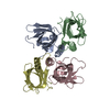
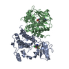
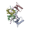
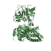
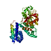
| Title | Crystal Structure of Methyltransferase MboIIa (Moraxella bovis) | ||||||
 Components Components | Adenine-specific Methyltransferase MboIIA | ||||||
 Keywords Keywords | TRANSFERASE / STRUCTURAL GENOMICS / MORAXELLA BOVIS / DNA METHYLATION / S-ADENOSYLMETHIONINE / PSI / Protein Structure Initiative / Midwest Center for Structural Genomics / MCSG | ||||||
| Function / homology |  Function and homology information Function and homology informationN-methyltransferase activity / site-specific DNA-methyltransferase (adenine-specific) / site-specific DNA-methyltransferase (adenine-specific) activity / DNA restriction-modification system / methylation / DNA binding / cytoplasm Similarity search - Function | ||||||
| Biological species |  Moraxella bovis (bacteria) Moraxella bovis (bacteria) | ||||||
| Method |  X-RAY DIFFRACTION / SYNCHROTRON / MAD / Resolution: 1.74 Å X-RAY DIFFRACTION / SYNCHROTRON / MAD / Resolution: 1.74 Å | ||||||
 Authors Authors | Osipiuk, J. / Walsh, M.A. / Joachimiak, A. / Midwest Center for Structural Genomics (MCSG) | ||||||
 Citation Citation | Journal: Nucleic Acids Res. / Year: 2003 Title: Crystal structure of MboIIA methyltransferase. Authors: Osipiuk, J. / Walsh, M.A. / Joachimiak, A. #1: Journal: Nucleic Acids Res. / Year: 1991Title: Cloning and Characterization of the MboII Restriction-modification System Authors: Bocklage, H. / Heeger, K. / Muller-Hill, B. | ||||||
| History |
| ||||||
| Remark 999 | SEQUENCE DNA SEQUENCING OF THE CLONES SHOWED THERE IS A ALANINE CODON FOR RESIDUE 51 AND A ARGININE ...SEQUENCE DNA SEQUENCING OF THE CLONES SHOWED THERE IS A ALANINE CODON FOR RESIDUE 51 AND A ARGININE CODON FOR RESIDUE 111. |
- Structure visualization
| Structure viewer | Molecule: MolmilJmol/JSmol |
|---|
- Downloads & links
-Download
| PDBx/mmCIF format | 1g60.cif.gz | 118.1 KB | Display | PDBx/mmCIF format |
|---|---|---|---|---|
| PDB format | pdb1g60.ent.gz | 91.2 KB | Display | PDB format |
| PDBx/mmJSON format | 1g60.json.gz | Tree view | PDBx/mmJSON format | |
| Others |  Other downloads Other downloads |
-Validation report
| Arichive directory | https://data.pdbj.org/pub/pdb/validation_reports/g6/1g60ftp://data.pdbj.org/pub/pdb/validation_reports/g6/1g60 | HTTPS FTP |
|---|
-Related structure data
| Similar structure data | |
|---|---|
| Other databases |

-Links
 PDBj
PDBj
- Assembly
| Deposited unit | 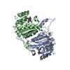
| ||||||||
|---|---|---|---|---|---|---|---|---|---|
| 1 |
| ||||||||
| Unit cell |
|
-Components
| #1: Protein | Mass: 30192.391 Da / Num. of mol.: 2 Source method: isolated from a genetically manipulated source Source: (gene. exp.) Moraxella bovis (bacteria) / Plasmid: PET24A / Production host: References: UniProt: P23192, site-specific DNA-methyltransferase (adenine-specific) #2: Chemical |   Mass: 22.990 Da / Num. of mol.: 2 / Source method: obtained synthetically / Formula: Na Mass: 22.990 Da / Num. of mol.: 2 / Source method: obtained synthetically / Formula: Na#3: Chemical |   Mass: 398.437 Da / Num. of mol.: 2 / Source method: obtained synthetically / Formula: C15H22N6O5S Mass: 398.437 Da / Num. of mol.: 2 / Source method: obtained synthetically / Formula: C15H22N6O5S#4: Water | ChemComp-HOH / |  Mass: 18.015 Da / Num. of mol.: 361 / Source method: isolated from a natural source / Formula: H2O Mass: 18.015 Da / Num. of mol.: 361 / Source method: isolated from a natural source / Formula: H2O |
|---|
-Experimental details
-Experiment
| Experiment | Method: X-RAY DIFFRACTION / Number of used crystals: 1 |
|---|
- Sample preparation
| Crystal | Density Matthews: 2.47 Å3/Da / Density % sol: 50.29 % | ||||||||||||||||||||||||||||||||||||||||||||||||||||||||||||||||||||||
|---|---|---|---|---|---|---|---|---|---|---|---|---|---|---|---|---|---|---|---|---|---|---|---|---|---|---|---|---|---|---|---|---|---|---|---|---|---|---|---|---|---|---|---|---|---|---|---|---|---|---|---|---|---|---|---|---|---|---|---|---|---|---|---|---|---|---|---|---|---|---|---|
| Crystal grow | Temperature: 277 K / Method: vapor diffusion, hanging drop / pH: 8.4 Details: 4% PEG monomethyl ether 5000, 50 mM potassium chloride, 50 mM sodium chloride, 1 mM DTT, 50 mM bicine, pH 8.4, VAPOR DIFFUSION, HANGING DROP, temperature 277.0K | ||||||||||||||||||||||||||||||||||||||||||||||||||||||||||||||||||||||
| Crystal grow | *PLUS Temperature: 277 K / pH: 7.5 | ||||||||||||||||||||||||||||||||||||||||||||||||||||||||||||||||||||||
| Components of the solutions | *PLUS
|
-Data collection
| Diffraction | Mean temperature: 100 K |
|---|---|
| Diffraction source | Source: SYNCHROTRON / Site: APS  / Beamline: 19-ID / Wavelength: 1.00599 Å / Beamline: 19-ID / Wavelength: 1.00599 Å |
| Detector | Type: CUSTOM-MADE / Detector: CCD / Date: Aug 29, 1999 |
| Radiation | Monochromator: SI 111 / Protocol: MAD / Monochromatic (M) / Laue (L): M / Scattering type: x-ray |
| Radiation wavelength | Wavelength: 1.00599 Å / Relative weight: 1 |
| Reflection | Resolution: 1.74→28.79 Å / Num. obs: 60668 / % possible obs: 99.7 % / Observed criterion σ(F): 1 / Observed criterion σ(I): 1 / Redundancy: 3.72 % / Biso Wilson estimate: 22 Å2 / Rmerge(I) obs: 0.066 / Net I/σ(I): 12.1 |
| Reflection shell | Resolution: 1.74→1.77 Å / Redundancy: 3.63 % / Rmerge(I) obs: 0.339 / Mean I/σ(I) obs: 3.4 / Num. unique all: 2974 / % possible all: 99.4 |
| Reflection | *PLUS Lowest resolution: 30 Å / Num. obs: 60068 |
| Reflection shell | *PLUS % possible obs: 99.4 % |
- Processing
| Software |
| |||||||||||||||||||||||||
|---|---|---|---|---|---|---|---|---|---|---|---|---|---|---|---|---|---|---|---|---|---|---|---|---|---|---|
| Refinement | Method to determine structure: MAD / Resolution: 1.74→29.75 Å / σ(F): 0 / σ(I): 0 / Stereochemistry target values: Engh & Huber
| |||||||||||||||||||||||||
| Displacement parameters | Biso mean: 27.7 Å2
| |||||||||||||||||||||||||
| Refine analyze |
| |||||||||||||||||||||||||
| Refinement step | Cycle: LAST / Resolution: 1.74→29.75 Å
| |||||||||||||||||||||||||
| Refine LS restraints |
| |||||||||||||||||||||||||
| LS refinement shell | Resolution: 1.74→1.826 Å
| |||||||||||||||||||||||||
| Refinement | *PLUS Lowest resolution: 30 Å / Rfactor obs: 0.198 | |||||||||||||||||||||||||
| Solvent computation | *PLUS | |||||||||||||||||||||||||
| Displacement parameters | *PLUS |
