 Movie
Movie Controller

[English] 日本語
 Yorodumi
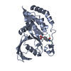
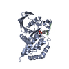
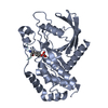
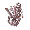
- PDB-4qum: Crystal structure of PTPN3 (PTPH1) in complex with a dually phosp... -
+ Open data

- Basic information
| Entry | Database: PDB / ID: 4qum | ||||||
|---|---|---|---|---|---|---|---|
| Title | Crystal structure of PTPN3 (PTPH1) in complex with a dually phosphorylated MAPK12 peptide | ||||||
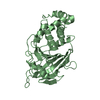
 Components Components |
| ||||||
 Keywords Keywords | HYDROLASE / Alpha Beta | ||||||
| Function / homology |  Function and homology information Function and homology informationregulation of membrane depolarization during action potential / negative regulation of membrane protein ectodomain proteolysis / regulation of sodium ion transmembrane transport / myoblast differentiation / negative regulation of mitotic cell cycle / Activation of PPARGC1A (PGC-1alpha) by phosphorylation / peptidase activator activity / positive regulation of muscle cell differentiation / muscle organ development / Myogenesis ...regulation of membrane depolarization during action potential / negative regulation of membrane protein ectodomain proteolysis / regulation of sodium ion transmembrane transport / myoblast differentiation / negative regulation of mitotic cell cycle / Activation of PPARGC1A (PGC-1alpha) by phosphorylation / peptidase activator activity / positive regulation of muscle cell differentiation / muscle organ development / Myogenesis / negative regulation of cell cycle / negative regulation of epidermal growth factor receptor signaling pathway / MAP kinase activity / mitogen-activated protein kinase / sodium channel regulator activity / cytoskeletal protein binding / signal transduction in response to DNA damage / phosphotyrosine residue binding / p38MAPK events / protein-tyrosine-phosphatase / protein tyrosine phosphatase activity / EGFR downregulation / NOD1/2 Signaling Pathway / VEGFA-VEGFR2 Pathway / cytoplasmic side of plasma membrane / Negative regulation of MAPK pathway / MAPK cascade / ATPase binding / cytoskeleton / regulation of cell cycle / intracellular signal transduction / protein serine kinase activity / protein serine/threonine kinase activity / magnesium ion binding / signal transduction / mitochondrion / nucleoplasm / ATP binding / nucleus / plasma membrane / cytoplasm / cytosol Similarity search - Function | ||||||
| Biological species |  Homo sapiens (human) Homo sapiens (human) | ||||||
| Method |  X-RAY DIFFRACTION / SYNCHROTRON / MOLECULAR REPLACEMENT / Resolution: 2.516 Å X-RAY DIFFRACTION / SYNCHROTRON / MOLECULAR REPLACEMENT / Resolution: 2.516 Å | ||||||
 Authors Authors | Chen, K.E. / Meng, T.C. / Wang, A.H.J. | ||||||
 Citation Citation | Journal: Sci.Signal. / Year: 2014 Title: Reciprocal allosteric regulation of p38 gamma and PTPN3 involves a PDZ domain-modulated complex formation. Authors: Chen, K.E. / Lin, S.Y. / Wu, M.J. / Ho, M.R. / Santhanam, A. / Chou, C.C. / Meng, T.C. / Wang, A.H.J. | ||||||
| History |
|
- Structure visualization
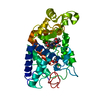
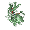
| Structure viewer | Molecule: MolmilJmol/JSmol |
|---|
- Downloads & links
-Download
| PDBx/mmCIF format | 4qum.cif.gz | 72.9 KB | Display | PDBx/mmCIF format |
|---|---|---|---|---|
| PDB format | pdb4qum.ent.gz | 52.9 KB | Display | PDB format |
| PDBx/mmJSON format | 4qum.json.gz | Tree view | PDBx/mmJSON format | |
| Others |  Other downloads Other downloads |
-Validation report
| Arichive directory | https://data.pdbj.org/pub/pdb/validation_reports/qu/4qumftp://data.pdbj.org/pub/pdb/validation_reports/qu/4qum | HTTPS FTP |
|---|
-Related structure data
| Related structure data |  4qunC  2b49S C: citing same article ( S: Starting model for refinement |
|---|---|
| Similar structure data |


-Links
 PDBj
PDBj


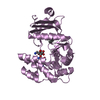
- Assembly
| Deposited unit | 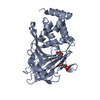
| ||||||||
|---|---|---|---|---|---|---|---|---|---|
| 1 |
| ||||||||
| Unit cell |
|
-Components
| #1: Protein | Mass: 34887.664 Da / Num. of mol.: 1 / Fragment: Catalytic domain (UNP RESIDUES 628-909) / Mutation: D811A, C842S Source method: isolated from a genetically manipulated source Source: (gene. exp.) Homo sapiens (human) / Gene: PTPH1, PTPN3 / Plasmid: pMCSG7 / Production host:  |
|---|---|
| #2: Protein/peptide | Mass: 1244.252 Da / Num. of mol.: 1 / Fragment: Activation loop (UNP RESIDUES 182-190) / Mutation: W190R / Source method: obtained synthetically / Source: (synth.) Homo sapiens (human)References: UniProt: P53778, mitogen-activated protein kinase |
| #3: Water | ChemComp-HOH /  Mass: 18.015 Da / Num. of mol.: 27 / Source method: isolated from a natural source / Formula: H2O Mass: 18.015 Da / Num. of mol.: 27 / Source method: isolated from a natural source / Formula: H2O |
| Has protein modification | Y |
-Experimental details
-Experiment
| Experiment | Method: X-RAY DIFFRACTION / Number of used crystals: 1 |
|---|
- Sample preparation
| Crystal | Density Matthews: 2.5 Å3/Da / Density % sol: 50.71 % |
|---|---|
| Crystal grow | Temperature: 293 K / Method: vapor diffusion, sitting drop / pH: 8.5 Details: 0.1M Tris-HCl, 30% PEG 8000, 5% glycerol, pH 8.5, VAPOR DIFFUSION, SITTING DROP, temperature 293K |
-Data collection
| Diffraction | Mean temperature: 100 K |
|---|---|
| Diffraction source | Source: SYNCHROTRON / Site: NSRRC  / Beamline: BL15A1 / Wavelength: 1 Å / Beamline: BL15A1 / Wavelength: 1 Å |
| Detector | Type: RAYONIX MX300HE / Detector: CCD / Date: Mar 6, 2013 |
| Radiation | Monochromator: Si 111 CHANNEL / Protocol: SINGLE WAVELENGTH / Monochromatic (M) / Laue (L): M / Scattering type: x-ray |
| Radiation wavelength | Wavelength: 1 Å / Relative weight: 1 |
| Reflection | Resolution: 2.51→50 Å / Num. all: 12505 / Num. obs: 12505 / % possible obs: 98 % / Observed criterion σ(F): 0 / Observed criterion σ(I): 0 / Redundancy: 4.7 % / Biso Wilson estimate: 41.1 Å2 / Rmerge(I) obs: 0.122 / Net I/σ(I): 10.79 |
| Reflection shell | Resolution: 2.51→2.6 Å / Redundancy: 4 % / Rmerge(I) obs: 0.521 / Mean I/σ(I) obs: 2.35 / Num. unique all: 1182 / % possible all: 95.1 |
- Processing
| Software |
| ||||||||||||||||||||||||||||||
|---|---|---|---|---|---|---|---|---|---|---|---|---|---|---|---|---|---|---|---|---|---|---|---|---|---|---|---|---|---|---|---|
| Refinement | Method to determine structure: MOLECULAR REPLACEMENT Starting model: PDB Entry 2B49 Resolution: 2.516→31.325 Å / SU ML: 0.34 / σ(F): 1.34 / Phase error: 25.22 / Stereochemistry target values: MLHL
| ||||||||||||||||||||||||||||||
| Solvent computation | Shrinkage radii: 0.9 Å / VDW probe radii: 1.11 Å / Solvent model: FLAT BULK SOLVENT MODEL | ||||||||||||||||||||||||||||||
| Refinement step | Cycle: LAST / Resolution: 2.516→31.325 Å
| ||||||||||||||||||||||||||||||
| Refine LS restraints |
| ||||||||||||||||||||||||||||||
| LS refinement shell | Refine-ID: X-RAY DIFFRACTION / Total num. of bins used: 4
|
