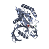
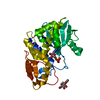
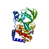
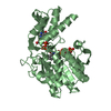
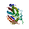
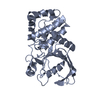
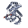
Entry Database : PDB / ID : 6l03Title structure of PTP-MEG2 and MUNC18-1-pY145 peptide complex Tyrosine-protein phosphatase non-receptor type 9 stxbp1-pY145 peptide Keywords / / / Function / homology Function Domain/homology Component
/ / / / / / / / / / / / / / / / / / / / / / / / / / / / / / / / / / / / / / / / / / / / / / / / / / / / / / / / / / / / / / / / / / / / / / / / / / / / / / / / / / / / / / / / / / / / / / / / / / / / / / / / / / / / / / / / / / / / / Biological species Homo sapiens (human)Method / / / Resolution : 2.084 Å Authors Xu, Y.F. / Chen, X. / Yu, X. / Sun, J.P. Journal : Embo Rep. / Year : 2021Title : PTP-MEG2 regulates quantal size and fusion pore opening through two distinct structural bases and substrates.Authors: Xu, Y.F. / Chen, X. / Yang, Z. / Xiao, P. / Liu, C.H. / Li, K.S. / Yang, X.Z. / Wang, Y.J. / Zhu, Z.L. / Xu, Z.G. / Zhang, S. / Wang, C. / Song, Y.C. / Zhao, W.D. / Wang, C.H. / Ji, Z.L. / ... Authors : Xu, Y.F. / Chen, X. / Yang, Z. / Xiao, P. / Liu, C.H. / Li, K.S. / Yang, X.Z. / Wang, Y.J. / Zhu, Z.L. / Xu, Z.G. / Zhang, S. / Wang, C. / Song, Y.C. / Zhao, W.D. / Wang, C.H. / Ji, Z.L. / Zhang, Z.Y. / Cui, M. / Sun, J.P. / Yu, X. History Deposition Sep 25, 2019 Deposition site / Processing site Revision 1.0 Sep 30, 2020 Provider / Type Revision 1.1 Oct 13, 2021 Group / Category / citation_author / database_2Item _citation.country / _citation.journal_abbrev ... _citation.country / _citation.journal_abbrev / _citation.journal_id_CSD / _citation.journal_id_ISSN / _citation.journal_volume / _citation.page_first / _citation.page_last / _citation.pdbx_database_id_DOI / _citation.pdbx_database_id_PubMed / _citation.title / _citation.year / _database_2.pdbx_DOI / _database_2.pdbx_database_accession Revision 1.2 Nov 22, 2023 Group / Refinement descriptionCategory / chem_comp_bond / pdbx_initial_refinement_modelRevision 1.3 Oct 23, 2024 Group / Category / pdbx_modification_feature / Item
Show all Show less
 Movie
Movie Controller


 Open data
 Basic information
 Components
 Keywords
 Function and homology information
 Homo sapiens (human)
 X-RAY DIFFRACTION /
  Authors
 Citation
 Structure visualization
 Downloads & links
 Other downloads
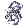


 PDBj
PDBj


 Assembly
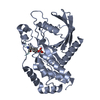

 Mass: 18.015 Da / Num. of mol.: 79 / Source method: isolated from a natural source / Formula: H2O
 Sample preparation
 / Beamline: BL17U1 / Wavelength: 0.9788 Å
 Processing