 Movie
Movie Controller

[English] 日本語
 Yorodumi
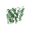
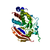
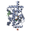
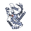
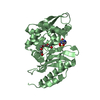
- PDB-4d2o: Crystal structure of the class A extended-spectrum beta-lactamase... -
+ Open data

- Basic information
| Entry | Database: PDB / ID: 4d2o | |||||||||
|---|---|---|---|---|---|---|---|---|---|---|
| Title | Crystal structure of the class A extended-spectrum beta-lactamase PER- 2 | |||||||||
 Components Components | PER-2 BETA-LACTAMASE | |||||||||
 Keywords Keywords | HYDROLASE / OXYIMINO-CEPHALOSPORINASE / ESBL / CEFOTAXIMASE | |||||||||
| Function / homology |  Function and homology information Function and homology informationbeta-lactam antibiotic catabolic process / beta-lactamase activity / beta-lactamase / response to antibiotic Similarity search - Function | |||||||||
| Biological species |  CITROBACTER FREUNDII (bacteria) CITROBACTER FREUNDII (bacteria) | |||||||||
| Method |  X-RAY DIFFRACTION / SYNCHROTRON / MOLECULAR REPLACEMENT / Resolution: 2.2 Å X-RAY DIFFRACTION / SYNCHROTRON / MOLECULAR REPLACEMENT / Resolution: 2.2 Å | |||||||||
 Authors Authors | Power, P. / Herman, R. / Ruggiero, M. / Kerff, F. / Galleni, M. / Gutkind, G. / Charlier, P. / Sauvage, E. | |||||||||
 Citation Citation | Journal: Antimicrob.Agents Chemother. / Year: 2014 Title: Crystal Structure of the Extended-Spectrum Beta-Lactamase Per- 2 and Insights Into the Role of Specific Residues in the Interaction with Beta-Lactams and Beta-Lactamase Inhibitors. Authors: Ruggiero, M. / Sauvage, E. / Herman, R. / Galleni, M. / Gutkind, G. / Charlier, P. / Power, P. | |||||||||
| History |
|
- Structure visualization
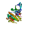
| Structure viewer | Molecule: MolmilJmol/JSmol |
|---|
- Downloads & links
-Download
| PDBx/mmCIF format | 4d2o.cif.gz | 219.8 KB | Display | PDBx/mmCIF format |
|---|---|---|---|---|
| PDB format | pdb4d2o.ent.gz | 177.9 KB | Display | PDB format |
| PDBx/mmJSON format | 4d2o.json.gz | Tree view | PDBx/mmJSON format | |
| Others |  Other downloads Other downloads |
-Validation report
| Arichive directory | https://data.pdbj.org/pub/pdb/validation_reports/d2/4d2oftp://data.pdbj.org/pub/pdb/validation_reports/d2/4d2o | HTTPS FTP |
|---|
-Related structure data
| Related structure data | 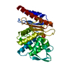 1e25S S: Starting model for refinement |
|---|---|
| Similar structure data |


-Links
 PDBj
PDBj


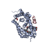
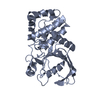
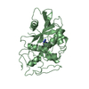
- Assembly
| Deposited unit | 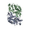
| ||||||||
|---|---|---|---|---|---|---|---|---|---|
| 1 | 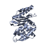
| ||||||||
| 2 | 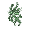
| ||||||||
| Unit cell |
|
-Components
| #1: Protein | Mass: 30956.328 Da / Num. of mol.: 2 / Fragment: RESIDUES 25-308 Source method: isolated from a genetically manipulated source Source: (gene. exp.) CITROBACTER FREUNDII (bacteria) / Production host: #2: Water | ChemComp-HOH / |  Mass: 18.015 Da / Num. of mol.: 152 / Source method: isolated from a natural source / Formula: H2O Mass: 18.015 Da / Num. of mol.: 152 / Source method: isolated from a natural source / Formula: H2O |
|---|
-Experimental details
-Experiment
| Experiment | Method: X-RAY DIFFRACTION / Number of used crystals: 1 |
|---|
- Sample preparation
| Crystal | Density Matthews: 1.89 Å3/Da / Density % sol: 34.3 % / Description: NONE |
|---|---|
| Crystal grow | Details: 0.1 M HEPES, 1.5 M SODIUM CITRATE BUFFER (PH 7.5). |
-Data collection
| Diffraction | Mean temperature: 100 K |
|---|---|
| Diffraction source | Source: SYNCHROTRON / Site: SOLEIL  / Beamline: PROXIMA 1 / Wavelength: 0.98011 / Beamline: PROXIMA 1 / Wavelength: 0.98011 |
| Detector | Type: DECTRIS PILATUS 6M / Detector: PIXEL / Date: Sep 23, 2012 |
| Radiation | Protocol: SINGLE WAVELENGTH / Monochromatic (M) / Laue (L): M / Scattering type: x-ray |
| Radiation wavelength | Wavelength: 0.98011 Å / Relative weight: 1 |
| Reflection | Resolution: 2.2→41.94 Å / Num. obs: 23354 / % possible obs: 100 % / Observed criterion σ(I): 0 / Redundancy: 6.8 % / Rmerge(I) obs: 0.14 / Net I/σ(I): 10.5 |
| Reflection shell | Resolution: 2.2→2.32 Å / Redundancy: 6.9 % / Rmerge(I) obs: 0.65 / Mean I/σ(I) obs: 3.1 / % possible all: 100 |
- Processing
| Software |
| ||||||||||||||||||||||||||||||||||||||||||||||||||||||||||||||||||||||||||||||||||||||||||||||||||||||||||||||||||||||||||||||||||||||||||||||||||||||||||||||||||||||||||||||||||||||
|---|---|---|---|---|---|---|---|---|---|---|---|---|---|---|---|---|---|---|---|---|---|---|---|---|---|---|---|---|---|---|---|---|---|---|---|---|---|---|---|---|---|---|---|---|---|---|---|---|---|---|---|---|---|---|---|---|---|---|---|---|---|---|---|---|---|---|---|---|---|---|---|---|---|---|---|---|---|---|---|---|---|---|---|---|---|---|---|---|---|---|---|---|---|---|---|---|---|---|---|---|---|---|---|---|---|---|---|---|---|---|---|---|---|---|---|---|---|---|---|---|---|---|---|---|---|---|---|---|---|---|---|---|---|---|---|---|---|---|---|---|---|---|---|---|---|---|---|---|---|---|---|---|---|---|---|---|---|---|---|---|---|---|---|---|---|---|---|---|---|---|---|---|---|---|---|---|---|---|---|---|---|---|---|
| Refinement | Method to determine structure: MOLECULAR REPLACEMENT Starting model: PDB ENTRY 1E25 Resolution: 2.2→33.46 Å / Cor.coef. Fo:Fc: 0.944 / Cor.coef. Fo:Fc free: 0.911 / SU B: 13.878 / SU ML: 0.172 / Cross valid method: THROUGHOUT / ESU R: 0.431 / ESU R Free: 0.24 / Stereochemistry target values: MAXIMUM LIKELIHOOD / Details: HYDROGENS HAVE BEEN ADDED IN THE RIDING POSITIONS.
| ||||||||||||||||||||||||||||||||||||||||||||||||||||||||||||||||||||||||||||||||||||||||||||||||||||||||||||||||||||||||||||||||||||||||||||||||||||||||||||||||||||||||||||||||||||||
| Solvent computation | Ion probe radii: 0.8 Å / Shrinkage radii: 0.8 Å / VDW probe radii: 1.2 Å / Solvent model: MASK | ||||||||||||||||||||||||||||||||||||||||||||||||||||||||||||||||||||||||||||||||||||||||||||||||||||||||||||||||||||||||||||||||||||||||||||||||||||||||||||||||||||||||||||||||||||||
| Displacement parameters | Biso mean: 29.24 Å2
| ||||||||||||||||||||||||||||||||||||||||||||||||||||||||||||||||||||||||||||||||||||||||||||||||||||||||||||||||||||||||||||||||||||||||||||||||||||||||||||||||||||||||||||||||||||||
| Refinement step | Cycle: LAST / Resolution: 2.2→33.46 Å
| ||||||||||||||||||||||||||||||||||||||||||||||||||||||||||||||||||||||||||||||||||||||||||||||||||||||||||||||||||||||||||||||||||||||||||||||||||||||||||||||||||||||||||||||||||||||
| Refine LS restraints |
|