 Movie
Movie Controller

[English] 日本語
 Yorodumi
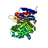
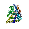
- PDB-6d3g: PER-2 class A extended-spectrum beta-lactamase crystal structure ... -
+ Open data

- Basic information
| Entry | Database: PDB / ID: 6d3g | |||||||||
|---|---|---|---|---|---|---|---|---|---|---|
| Title | PER-2 class A extended-spectrum beta-lactamase crystal structure in complex with avibactam at 2.4 Angstrom resolution | |||||||||
 Components Components | Beta-lactamase | |||||||||
 Keywords Keywords | HYDROLASE/HYDROLASE INHIBITOR / DBO / penicilloil-serine-transferase / antibiotic resistance / HYDROLASE / HYDROLASE-HYDROLASE INHIBITOR complex | |||||||||
| Function / homology |  Function and homology information Function and homology informationbeta-lactam antibiotic catabolic process / beta-lactamase activity / beta-lactamase / response to antibiotic Similarity search - Function | |||||||||
| Biological species |  Citrobacter freundii (bacteria) Citrobacter freundii (bacteria) | |||||||||
| Method |  X-RAY DIFFRACTION / SYNCHROTRON / MOLECULAR REPLACEMENT / Resolution: 2.398 Å X-RAY DIFFRACTION / SYNCHROTRON / MOLECULAR REPLACEMENT / Resolution: 2.398 Å | |||||||||
 Authors Authors | Power, P. / Ruggiero, M. / Gutkind, G. / Bonomo, R. / Klinke, S. | |||||||||
| Funding support |  Argentina, 2items Argentina, 2items
| |||||||||
 Citation Citation | Journal: Antimicrob.Agents Chemother. / Year: 2019 Title: Structural Insights into the Inhibition of the Extended-Spectrum beta-Lactamase PER-2 by Avibactam. Authors: Ruggiero, M. / Papp-Wallace, K.M. / Brunetti, F. / Barnes, M.D. / Bonomo, R.A. / Gutkind, G. / Klinke, S. / Power, P. | |||||||||
| History |
|
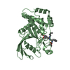
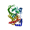
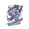
- Structure visualization
| Structure viewer | Molecule: MolmilJmol/JSmol |
|---|
- Downloads & links
-Download
| PDBx/mmCIF format | 6d3g.cif.gz | 222.9 KB | Display | PDBx/mmCIF format |
|---|---|---|---|---|
| PDB format | pdb6d3g.ent.gz | 180.2 KB | Display | PDB format |
| PDBx/mmJSON format | 6d3g.json.gz | Tree view | PDBx/mmJSON format | |
| Others |  Other downloads Other downloads |
-Validation report
| Arichive directory | https://data.pdbj.org/pub/pdb/validation_reports/d3/6d3gftp://data.pdbj.org/pub/pdb/validation_reports/d3/6d3g | HTTPS FTP |
|---|
-Related structure data
| Related structure data | 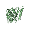 6dguC 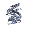 4d2oS S: Starting model for refinement C: citing same article ( |
|---|---|
| Similar structure data |


-Links
 PDBj
PDBj


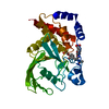
- Assembly
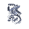
| Deposited unit | 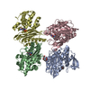
| ||||||||
|---|---|---|---|---|---|---|---|---|---|
| 1 | 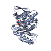
| ||||||||
| 2 | 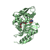
| ||||||||
| 3 | 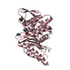
| ||||||||
| 4 | 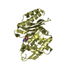
| ||||||||
| Unit cell |
|
-Components
| #1: Protein | Mass: 30956.328 Da / Num. of mol.: 4 Source method: isolated from a genetically manipulated source Source: (gene. exp.) Citrobacter freundii (bacteria) / Gene: blaPER-2 / Plasmid: pET28/PER-2 / Production host: #2: Chemical | ChemComp-NXL / ( 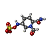  Mass: 267.260 Da / Num. of mol.: 4 / Source method: obtained synthetically / Formula: C7H13N3O6S / Comment: antibiotic, inhibitor*YM Mass: 267.260 Da / Num. of mol.: 4 / Source method: obtained synthetically / Formula: C7H13N3O6S / Comment: antibiotic, inhibitor*YM#3: Chemical | ChemComp-PG4 / |   Mass: 194.226 Da / Num. of mol.: 1 / Source method: obtained synthetically / Formula: C8H18O5 / Comment: precipitant*YM Mass: 194.226 Da / Num. of mol.: 1 / Source method: obtained synthetically / Formula: C8H18O5 / Comment: precipitant*YM#4: Water | ChemComp-HOH / |  Mass: 18.015 Da / Num. of mol.: 74 / Source method: isolated from a natural source / Formula: H2O Mass: 18.015 Da / Num. of mol.: 74 / Source method: isolated from a natural source / Formula: H2OHas protein modification | Y | |
|---|
-Experimental details
-Experiment
| Experiment | Method: X-RAY DIFFRACTION / Number of used crystals: 1 |
|---|
- Sample preparation
| Crystal | Density Matthews: 2.35 Å3/Da / Density % sol: 47.72 % |
|---|---|
| Crystal grow | Temperature: 294 K / Method: vapor diffusion, hanging drop / Details: 20% PEG 6000, 0.05 M imidazole pH=8.0 |
-Data collection
| Diffraction | Mean temperature: 100 K |
|---|---|
| Diffraction source | Source: SYNCHROTRON / Site: SOLEIL  / Beamline: PROXIMA 2 / Wavelength: 0.9801 Å / Beamline: PROXIMA 2 / Wavelength: 0.9801 Å |
| Detector | Type: DECTRIS EIGER X 9M / Detector: PIXEL / Date: Dec 11, 2016 Details: CONVEX PREFOCUSSING MIRROR AND A KIRKPATRICK-BAEZ PAIR OF FOCUSSING MIRRORS |
| Radiation | Monochromator: CRYOGENICALLY COOLED CHANNEL CUT SI[111] CRYSTAL MONOCHROMATOR Protocol: SINGLE WAVELENGTH / Monochromatic (M) / Laue (L): M / Scattering type: x-ray |
| Radiation wavelength | Wavelength: 0.9801 Å / Relative weight: 1 |
| Reflection | Resolution: 2.398→48.17 Å / Num. obs: 46410 / % possible obs: 99.76 % / Redundancy: 6.65 % / Biso Wilson estimate: 55.8 Å2 / CC1/2: 0.999 / Rmerge(I) obs: 0.085 / Rrim(I) all: 0.092 / Net I/σ(I): 13.73 |
| Reflection shell | Resolution: 2.4→2.54 Å / Redundancy: 6.65 % / Rmerge(I) obs: 0.9 / Mean I/σ(I) obs: 1.64 / Num. unique obs: 7289 / CC1/2: 0.746 / Rrim(I) all: 1 / % possible all: 98.8 |
- Processing
| Software |
| ||||||||||||||||||||||||||||||||||||||||||||||||||||||||||||||||||||||||||||||||||||||||||||||||||||||||||||||||||||||||||||||
|---|---|---|---|---|---|---|---|---|---|---|---|---|---|---|---|---|---|---|---|---|---|---|---|---|---|---|---|---|---|---|---|---|---|---|---|---|---|---|---|---|---|---|---|---|---|---|---|---|---|---|---|---|---|---|---|---|---|---|---|---|---|---|---|---|---|---|---|---|---|---|---|---|---|---|---|---|---|---|---|---|---|---|---|---|---|---|---|---|---|---|---|---|---|---|---|---|---|---|---|---|---|---|---|---|---|---|---|---|---|---|---|---|---|---|---|---|---|---|---|---|---|---|---|---|---|---|---|
| Refinement | Method to determine structure: MOLECULAR REPLACEMENT Starting model: 4D2O Resolution: 2.398→48.17 Å / SU ML: 0.35 / Cross valid method: THROUGHOUT / σ(F): 1.37 / Phase error: 28.31
| ||||||||||||||||||||||||||||||||||||||||||||||||||||||||||||||||||||||||||||||||||||||||||||||||||||||||||||||||||||||||||||||
| Solvent computation | Shrinkage radii: 0.9 Å / VDW probe radii: 1.11 Å | ||||||||||||||||||||||||||||||||||||||||||||||||||||||||||||||||||||||||||||||||||||||||||||||||||||||||||||||||||||||||||||||
| Displacement parameters | Biso mean: 65.73 Å2 | ||||||||||||||||||||||||||||||||||||||||||||||||||||||||||||||||||||||||||||||||||||||||||||||||||||||||||||||||||||||||||||||
| Refinement step | Cycle: LAST / Resolution: 2.398→48.17 Å
| ||||||||||||||||||||||||||||||||||||||||||||||||||||||||||||||||||||||||||||||||||||||||||||||||||||||||||||||||||||||||||||||
| Refine LS restraints |
| ||||||||||||||||||||||||||||||||||||||||||||||||||||||||||||||||||||||||||||||||||||||||||||||||||||||||||||||||||||||||||||||
| LS refinement shell |
|
