 Movie
Movie Controller

[English] 日本語
 Yorodumi
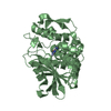
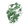
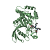
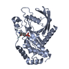
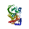
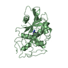
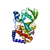
- PDB-1rpn: Crystal Structure of GDP-D-mannose 4,6-dehydratase in complexes w... -
+ Open data

- Basic information
| Entry | Database: PDB / ID: 1rpn | ||||||
|---|---|---|---|---|---|---|---|
| Title | Crystal Structure of GDP-D-mannose 4,6-dehydratase in complexes with GDP and NADPH | ||||||
 Components Components | GDP-mannose 4,6-dehydratase | ||||||
 Keywords Keywords | LYASE / short-chain dehydrogenase/reductase / Rossmann fold | ||||||
| Function / homology |  Function and homology information Function and homology informationGDP-D-rhamnose biosynthetic process / GDP-mannose 4,6-dehydratase / GDP-mannose 4,6-dehydratase activity / 'de novo' GDP-L-fucose biosynthetic process / O antigen biosynthetic process / lipopolysaccharide biosynthetic process / NADP+ binding Similarity search - Function | ||||||
| Biological species |   Pseudomonas aeruginosa (bacteria) Pseudomonas aeruginosa (bacteria) | ||||||
| Method |  X-RAY DIFFRACTION / SYNCHROTRON / MOLECULAR REPLACEMENT / Resolution: 2.15 Å X-RAY DIFFRACTION / SYNCHROTRON / MOLECULAR REPLACEMENT / Resolution: 2.15 Å | ||||||
 Authors Authors | Webb, N.A. / Mulichak, A.M. / Lam, J.S. / Rocchetta, H.L. / Garavito, R.M. | ||||||
 Citation Citation | Journal: Protein Sci. / Year: 2004 Title: Crystal structure of a tetrameric GDP-D-mannose 4,6-dehydratase from a bacterial GDP-D-rhamnose biosynthetic pathway. Authors: Webb, N.A. / Mulichak, A.M. / Lam, J.S. / Rocchetta, H.L. / Garavito, R.M. | ||||||
| History |
|
- Structure visualization
| Structure viewer | Molecule: MolmilJmol/JSmol |
|---|
- Downloads & links
-Download
| PDBx/mmCIF format | 1rpn.cif.gz | 279.9 KB | Display | PDBx/mmCIF format |
|---|---|---|---|---|
| PDB format | pdb1rpn.ent.gz | 224 KB | Display | PDB format |
| PDBx/mmJSON format | 1rpn.json.gz | Tree view | PDBx/mmJSON format | |
| Others |  Other downloads Other downloads |
-Validation report
| Arichive directory | https://data.pdbj.org/pub/pdb/validation_reports/rp/1rpnftp://data.pdbj.org/pub/pdb/validation_reports/rp/1rpn | HTTPS FTP |
|---|
-Related structure data
| Related structure data |  1n7hS S: Starting model for refinement |
|---|---|
| Similar structure data |


-Links
 PDBj
PDBj

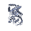
- Assembly
| Deposited unit | 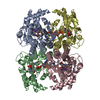
| ||||||||
|---|---|---|---|---|---|---|---|---|---|
| 1 | 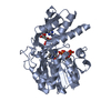
| ||||||||
| 2 | 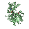
| ||||||||
| 3 | 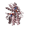
| ||||||||
| 4 | 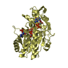
| ||||||||
| 5 |
| ||||||||
| Unit cell |
|
-Components
| #1: Protein | Mass: 37849.805 Da / Num. of mol.: 4 Source method: isolated from a genetically manipulated source Source: (gene. exp.) Pseudomonas aeruginosa (bacteria) / Gene: GCA, PA5453 / Plasmid: pQE30 / Production host: #2: Chemical | ChemComp-NDP /   Mass: 745.421 Da / Num. of mol.: 4 / Source method: obtained synthetically / Formula: C21H30N7O17P3 Mass: 745.421 Da / Num. of mol.: 4 / Source method: obtained synthetically / Formula: C21H30N7O17P3#3: Chemical | ChemComp-GDP /   Type: RNA linking / Mass: 443.201 Da / Num. of mol.: 4 / Source method: obtained synthetically / Formula: C10H15N5O11P2 / Comment: GDP, energy-carrying molecule*YM Type: RNA linking / Mass: 443.201 Da / Num. of mol.: 4 / Source method: obtained synthetically / Formula: C10H15N5O11P2 / Comment: GDP, energy-carrying molecule*YM#4: Water | ChemComp-HOH / |  Mass: 18.015 Da / Num. of mol.: 693 / Source method: isolated from a natural source / Formula: H2O Mass: 18.015 Da / Num. of mol.: 693 / Source method: isolated from a natural source / Formula: H2O |
|---|
-Experimental details
-Experiment
| Experiment | Method: X-RAY DIFFRACTION / Number of used crystals: 1 |
|---|
- Sample preparation
| Crystal | Density Matthews: 3.31 Å3/Da / Density % sol: 62.9 % | |||||||||||||||||||||||||||||||||||||||||||||||||
|---|---|---|---|---|---|---|---|---|---|---|---|---|---|---|---|---|---|---|---|---|---|---|---|---|---|---|---|---|---|---|---|---|---|---|---|---|---|---|---|---|---|---|---|---|---|---|---|---|---|---|
| Crystal grow | Temperature: 298 K / Method: vapor diffusion, hanging drop / pH: 8.5 Details: MPD, Tris, ammonium phosphate, pH 8.5, VAPOR DIFFUSION, HANGING DROP, temperature 298K | |||||||||||||||||||||||||||||||||||||||||||||||||
| Crystal grow | *PLUS Method: vapor diffusion, hanging drop | |||||||||||||||||||||||||||||||||||||||||||||||||
| Components of the solutions | *PLUS
|
-Data collection
| Diffraction | Mean temperature: 100 K |
|---|---|
| Diffraction source | Source: SYNCHROTRON / Site: APS  / Beamline: 5ID-B / Wavelength: 1 Å / Beamline: 5ID-B / Wavelength: 1 Å |
| Detector | Type: MARRESEARCH / Detector: CCD / Date: Apr 13, 2002 |
| Radiation | Protocol: SINGLE WAVELENGTH / Monochromatic (M) / Laue (L): M / Scattering type: x-ray |
| Radiation wavelength | Wavelength: 1 Å / Relative weight: 1 |
| Reflection | Resolution: 2.15→30 Å / Num. all: 110070 / Num. obs: 108095 / % possible obs: 98.3 % / Observed criterion σ(F): 0 / Observed criterion σ(I): -3 / Redundancy: 5 % / Rsym value: 0.039 / Net I/σ(I): 20 |
| Reflection shell | Resolution: 2.15→2.23 Å / Redundancy: 4 % / Num. unique all: 10063 / Rsym value: 0.079 / % possible all: 92.8 |
| Reflection | *PLUS Highest resolution: 2.2 Å / Rmerge(I) obs: 0.039 |
| Reflection shell | *PLUS % possible obs: 92.8 % / Rmerge(I) obs: 0.076 |
- Processing
| Software |
| |||||||||||||||||||||||||
|---|---|---|---|---|---|---|---|---|---|---|---|---|---|---|---|---|---|---|---|---|---|---|---|---|---|---|
| Refinement | Method to determine structure: MOLECULAR REPLACEMENT Starting model: PDB ENTRY 1N7H Resolution: 2.15→30 Å / Cross valid method: THROUGHOUT / σ(F): 1 / Stereochemistry target values: Engh & Huber
| |||||||||||||||||||||||||
| Displacement parameters | Biso mean: 20.8 Å2
| |||||||||||||||||||||||||
| Refine analyze |
| |||||||||||||||||||||||||
| Refinement step | Cycle: LAST / Resolution: 2.15→30 Å
| |||||||||||||||||||||||||
| Refine LS restraints |
| |||||||||||||||||||||||||
| Refinement | *PLUS Highest resolution: 2.2 Å / Num. reflection obs: 110170 / % reflection Rfree: 4.9 % / Rfactor Rfree: 0.195 / Rfactor Rwork: 0.173 | |||||||||||||||||||||||||
| Solvent computation | *PLUS | |||||||||||||||||||||||||
| Displacement parameters | *PLUS | |||||||||||||||||||||||||
| Refine LS restraints | *PLUS
|
