 Movie
Movie Controller

[English] 日本語
 Yorodumi
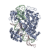
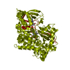
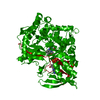
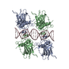
- PDB-1n7h: Crystal Structure of GDP-mannose 4,6-dehydratase ternary complex ... -
+ Open data

- Basic information
| Entry | Database: PDB / ID: 1n7h | ||||||
|---|---|---|---|---|---|---|---|
| Title | Crystal Structure of GDP-mannose 4,6-dehydratase ternary complex with NADPH and GDP | ||||||
 Components Components | GDP-D-mannose-4,6-dehydratase | ||||||
 Keywords Keywords | LYASE / Rossmann fold / SDR / short-chain dehydrogenase/reductase | ||||||
| Function / homology |  Function and homology information Function and homology informationGDP-mannose 4,6-dehydratase / GDP-mannose 4,6-dehydratase activity / unidimensional cell growth / 'de novo' GDP-L-fucose biosynthetic process / GTP binding / cytosol Similarity search - Function | ||||||
| Biological species |  | ||||||
| Method |  X-RAY DIFFRACTION / SYNCHROTRON / MOLECULAR REPLACEMENT / Resolution: 1.8 Å X-RAY DIFFRACTION / SYNCHROTRON / MOLECULAR REPLACEMENT / Resolution: 1.8 Å | ||||||
 Authors Authors | Mulichak, A.M. / Bonin, C.P. / Reiter, W.-D. / Garavito, R.M. | ||||||
 Citation Citation | Journal: Biochemistry / Year: 2002 Title: The structure of the MUR1 GDP-mannose 4,6-dehydratase from A. thaliana: Implications for ligand binding and specificity. Authors: Mulichak, A.M. / Bonin, C.P. / Reiter, W.-D. / Garavito, R.M. | ||||||
| History |
|
- Structure visualization
| Structure viewer | Molecule: MolmilJmol/JSmol |
|---|
- Downloads & links
-Download
| PDBx/mmCIF format | 1n7h.cif.gz | 156.8 KB | Display | PDBx/mmCIF format |
|---|---|---|---|---|
| PDB format | pdb1n7h.ent.gz | 121.9 KB | Display | PDB format |
| PDBx/mmJSON format | 1n7h.json.gz | Tree view | PDBx/mmJSON format | |
| Others |  Other downloads Other downloads |
-Validation report
| Arichive directory | https://data.pdbj.org/pub/pdb/validation_reports/n7/1n7hftp://data.pdbj.org/pub/pdb/validation_reports/n7/1n7h | HTTPS FTP |
|---|
-Related structure data


-Links
 PDBj
PDBj

- Assembly
| Deposited unit | 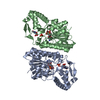
| |||||||||
|---|---|---|---|---|---|---|---|---|---|---|
| 1 | 
| |||||||||
| Unit cell |
| |||||||||
| Components on special symmetry positions |
| |||||||||
| Details | The biological assembly is a tetramer generated from the dimer in the asymmetric unit by the transformation: -x+1, y, -z+1/2 |
-Components
| #1: Protein | Mass: 43084.305 Da / Num. of mol.: 2 Source method: isolated from a genetically manipulated source Source: (gene. exp.)  #2: Chemical |   Mass: 745.421 Da / Num. of mol.: 2 / Source method: obtained synthetically / Formula: C21H30N7O17P3 Mass: 745.421 Da / Num. of mol.: 2 / Source method: obtained synthetically / Formula: C21H30N7O17P3#3: Chemical |   Type: RNA linking / Mass: 443.201 Da / Num. of mol.: 2 / Source method: obtained synthetically / Formula: C10H15N5O11P2 / Comment: GDP, energy-carrying molecule*YM Type: RNA linking / Mass: 443.201 Da / Num. of mol.: 2 / Source method: obtained synthetically / Formula: C10H15N5O11P2 / Comment: GDP, energy-carrying molecule*YM#4: Water | ChemComp-HOH / |  Mass: 18.015 Da / Num. of mol.: 440 / Source method: isolated from a natural source / Formula: H2O Mass: 18.015 Da / Num. of mol.: 440 / Source method: isolated from a natural source / Formula: H2O |
|---|
-Experimental details
-Experiment
| Experiment | Method: X-RAY DIFFRACTION / Number of used crystals: 1 |
|---|
- Sample preparation
| Crystal | Density Matthews: 2.45 Å3/Da / Density % sol: 49.42 % | |||||||||||||||
|---|---|---|---|---|---|---|---|---|---|---|---|---|---|---|---|---|
| Crystal grow | Temperature: 298 K / Method: vapor diffusion, hanging drop / pH: 6.4 Details: ammonium sulfate, PEG 400, Imidazole buffer, pH 6.4, VAPOR DIFFUSION, HANGING DROP, temperature 298K | |||||||||||||||
| Crystal grow | *PLUS | |||||||||||||||
| Components of the solutions | *PLUS
|
-Data collection
| Diffraction | Mean temperature: 100 K |
|---|---|
| Diffraction source | Source: SYNCHROTRON / Site: NSLS  / Beamline: X25 / Wavelength: 1 Å / Beamline: X25 / Wavelength: 1 Å |
| Detector | Type: BRANDEIS - B4 / Detector: CCD / Date: Oct 10, 2000 |
| Radiation | Protocol: SINGLE WAVELENGTH / Monochromatic (M) / Laue (L): M / Scattering type: x-ray |
| Radiation wavelength | Wavelength: 1 Å / Relative weight: 1 |
| Reflection | Resolution: 1.8→10 Å / Num. all: 70812 / Num. obs: 70104 / % possible obs: 95 % / Observed criterion σ(F): 0 / Observed criterion σ(I): 0 / Redundancy: 4 % / Rsym value: 0.063 |
| Reflection shell | Resolution: 1.8→1.86 Å / Redundancy: 4 % / Num. unique all: 6532 / Rsym value: 0.077 / % possible all: 88 |
| Reflection | *PLUS Lowest resolution: 10 Å / Num. obs: 70780 / % possible obs: 95 % / Rmerge(I) obs: 0.062 |
| Reflection shell | *PLUS % possible obs: 88 % / Rmerge(I) obs: 0.077 |
- Processing
| Software |
| |||||||||||||||||||||||||
|---|---|---|---|---|---|---|---|---|---|---|---|---|---|---|---|---|---|---|---|---|---|---|---|---|---|---|
| Refinement | Method to determine structure: MOLECULAR REPLACEMENT / Resolution: 1.8→10 Å / σ(F): 0 / Stereochemistry target values: Engh & Huber Details: Partial or no side chain atoms were refined for residues listed in REMARK 470.
| |||||||||||||||||||||||||
| Refine analyze | Luzzati coordinate error obs: 0.18 Å | |||||||||||||||||||||||||
| Refinement step | Cycle: LAST / Resolution: 1.8→10 Å
| |||||||||||||||||||||||||
| Refine LS restraints |
| |||||||||||||||||||||||||
| LS refinement shell | Resolution: 1.8→1.86 Å
| |||||||||||||||||||||||||
| Refinement | *PLUS Lowest resolution: 10 Å / % reflection Rfree: 5 % / Rfactor Rfree: 0.201 / Rfactor Rwork: 0.18 | |||||||||||||||||||||||||
| Solvent computation | *PLUS | |||||||||||||||||||||||||
| Displacement parameters | *PLUS | |||||||||||||||||||||||||
| Refine LS restraints | *PLUS
|
