- PDB-6kzq: structure of PTP-MEG2 and NSF-pY83 peptide complex -
+
Open data
ID or keywords:
Loading...
-
Basic information
Entry
Database: PDB / ID: 6kzq
Title
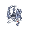
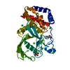
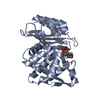
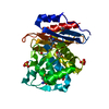
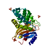
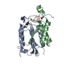
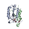
structure of PTP-MEG2 and NSF-pY83 peptide complex
Components
NSF-pY83 peptide
Tyrosine-protein phosphatase non-receptor type 9
Keywords
HYDROLASE / PTP-MEG2 / NSF / vesicle fusion
Function / homology
Function and homology information
: / Retrograde transport at the Trans-Golgi-Network / Trafficking of GluR2-containing AMPA receptors / Intra-Golgi traffic / SNARE complex disassembly / regulation of exocytosis / ATP-dependent protein disaggregase activity / intra-Golgi vesicle-mediated transport / Golgi to plasma membrane protein transport / Golgi stack ...: / Retrograde transport at the Trans-Golgi-Network / Trafficking of GluR2-containing AMPA receptors / Intra-Golgi traffic / SNARE complex disassembly / regulation of exocytosis / ATP-dependent protein disaggregase activity / intra-Golgi vesicle-mediated transport / Golgi to plasma membrane protein transport / Golgi stack / Interleukin-37 signaling / COPII-mediated vesicle transport / syntaxin-1 binding / protein dephosphorylation / vesicle-fusing ATPase / COPI-dependent Golgi-to-ER retrograde traffic / neuron projection terminus / exocytosis / peptidyl-tyrosine dephosphorylation / non-membrane spanning protein tyrosine phosphatase activity / positive regulation of receptor recycling / COPI-mediated anterograde transport / vesicle-mediated transport / protein-tyrosine-phosphatase / protein tyrosine phosphatase activity / ionotropic glutamate receptor binding / dendritic shaft / positive regulation of protein localization to plasma membrane / SNARE binding / PDZ domain binding / potassium ion transport / intracellular protein transport / positive regulation of protein catabolic process / negative regulation of neuron projection development / postsynaptic density / lysosomal membrane / protein kinase binding / protein-containing complex binding / Golgi apparatus / signal transduction / ATP hydrolysis activity / nucleoplasm / ATP binding / metal ion binding / plasma membrane / cytoplasm / cytosol Similarity search - Function
CRAL/TRIO, N-terminal domain / CRAL/TRIO, N-terminal domain / CRAL/TRIO, N-terminal domain superfamily / CRAL/TRIO domain / : / NSF, AAA+ ATPase lid domain / : / Vesicle-fusing ATPase / CRAL-TRIO lipid binding domain profile. / Domain in homologues of a S. cerevisiae phosphatidylinositol transfer protein (Sec14p) ...CRAL/TRIO, N-terminal domain / CRAL/TRIO, N-terminal domain / CRAL/TRIO, N-terminal domain superfamily / CRAL/TRIO domain / : / NSF, AAA+ ATPase lid domain / : / Vesicle-fusing ATPase / CRAL-TRIO lipid binding domain profile. / Domain in homologues of a S. cerevisiae phosphatidylinositol transfer protein (Sec14p) / CRAL-TRIO lipid binding domain / CRAL-TRIO lipid binding domain superfamily / Cell division protein 48 (CDC48), N-terminal domain / CDC48, N-terminal subdomain / Cell division protein 48 (CDC48) N-terminal domain / CDC48, domain 2 / Cell division protein 48 (CDC48), domain 2 / Cell division protein 48 (CDC48) domain 2 / CDC48 domain 2-like superfamily / Aspartate decarboxylase-like domain superfamily / Protein tyrosine phosphatase, catalytic domain / PTP type protein phosphatase domain profile. / Protein-tyrosine phosphatase / Tyrosine-specific protein phosphatase, PTPase domain / Protein-tyrosine phosphatase, catalytic / Protein tyrosine phosphatase, catalytic domain motif / Tyrosine specific protein phosphatases active site. / Protein-tyrosine phosphatase, active site / Tyrosine specific protein phosphatases domain profile. / Tyrosine-specific protein phosphatases domain / Protein-tyrosine phosphatase-like / AAA ATPase, AAA+ lid domain / AAA+ lid domain / ATPase, AAA-type, conserved site / AAA-protein family signature. / ATPase family associated with various cellular activities (AAA) / ATPase, AAA-type, core / ATPases associated with a variety of cellular activities / AAA+ ATPase domain / P-loop containing nucleoside triphosphate hydrolase Similarity search - Domain/homology
In the structure databanks used in Yorodumi, some data are registered as the other names, "COVID-19 virus" and "2019-nCoV". Here are the details of the virus and the list of structure data.
Jan 31, 2019. EMDB accession codes are about to change! (news from PDBe EMDB page)
EMDB accession codes are about to change! (news from PDBe EMDB page)
The allocation of 4 digits for EMDB accession codes will soon come to an end. Whilst these codes will remain in use, new EMDB accession codes will include an additional digit and will expand incrementally as the available range of codes is exhausted. The current 4-digit format prefixed with “EMD-” (i.e. EMD-XXXX) will advance to a 5-digit format (i.e. EMD-XXXXX), and so on. It is currently estimated that the 4-digit codes will be depleted around Spring 2019, at which point the 5-digit format will come into force.
The EM Navigator/Yorodumi systems omit the EMD- prefix.
Related info.:Q: What is EMD? / ID/Accession-code notation in Yorodumi/EM Navigator
Yorodumi is a browser for structure data from EMDB, PDB, SASBDB, etc.
This page is also the successor to EM Navigator detail page, and also detail information page/front-end page for Omokage search.
The word "yorodu" (or yorozu) is an old Japanese word meaning "ten thousand". "mi" (miru) is to see.
Related info.:EMDB / PDB / SASBDB / Comparison of 3 databanks / Yorodumi Search / Aug 31, 2016. New EM Navigator & Yorodumi / Yorodumi Papers / Jmol/JSmol / Function and homology information / Changes in new EM Navigator and Yorodumi
 Movie
Movie Controller


 Open data
 Basic information
 Components
 Keywords
 Function and homology information
 Homo sapiens (human)
 X-RAY DIFFRACTION /
  Authors
 Citation
 Structure visualization
 Downloads & links
 Other downloads
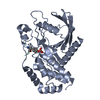


 PDBj
PDBj


 Assembly
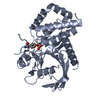

 Mass: 18.015 Da / Num. of mol.: 215 / Source method: isolated from a natural source / Formula: H2O
 Sample preparation
 / Beamline: BL17U1 / Wavelength: 0.9788 Å
 Processing