 Movie
Movie Controller

[English] 日本語
 Yorodumi
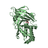
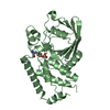
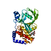
- PDB-4qun: Crystal structure of the PTPN3 (PTPH1) catalytic domain C842S mutant -
+ Open data

- Basic information
| Entry | Database: PDB / ID: 4qun | ||||||
|---|---|---|---|---|---|---|---|
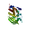
| Title | Crystal structure of the PTPN3 (PTPH1) catalytic domain C842S mutant | ||||||
 Components Components | Tyrosine-protein phosphatase non-receptor type 3 | ||||||
 Keywords Keywords | HYDROLASE / Alpha Beta | ||||||
| Function / homology |  Function and homology information Function and homology informationregulation of membrane depolarization during action potential / negative regulation of membrane protein ectodomain proteolysis / regulation of sodium ion transmembrane transport / negative regulation of mitotic cell cycle / negative regulation of epidermal growth factor receptor signaling pathway / sodium channel regulator activity / cytoskeletal protein binding / phosphotyrosine residue binding / protein-tyrosine-phosphatase / protein tyrosine phosphatase activity ...regulation of membrane depolarization during action potential / negative regulation of membrane protein ectodomain proteolysis / regulation of sodium ion transmembrane transport / negative regulation of mitotic cell cycle / negative regulation of epidermal growth factor receptor signaling pathway / sodium channel regulator activity / cytoskeletal protein binding / phosphotyrosine residue binding / protein-tyrosine-phosphatase / protein tyrosine phosphatase activity / EGFR downregulation / cytoplasmic side of plasma membrane / Negative regulation of MAPK pathway / MAPK cascade / ATPase binding / cytoskeleton / plasma membrane / cytoplasm / cytosol Similarity search - Function | ||||||
| Biological species |  Homo sapiens (human) Homo sapiens (human) | ||||||
| Method |  X-RAY DIFFRACTION / MOLECULAR REPLACEMENT / Resolution: 1.86 Å X-RAY DIFFRACTION / MOLECULAR REPLACEMENT / Resolution: 1.86 Å | ||||||
 Authors Authors | Chen, K.E. / Meng, T.C. / Wang, A.H.J. | ||||||
 Citation Citation | Journal: Sci.Signal. / Year: 2014 Title: Reciprocal allosteric regulation of p38 gamma and PTPN3 involves a PDZ domain-modulated complex formation. Authors: Chen, K.E. / Lin, S.Y. / Wu, M.J. / Ho, M.R. / Santhanam, A. / Chou, C.C. / Meng, T.C. / Wang, A.H.J. | ||||||
| History |
|
- Structure visualization
| Structure viewer | Molecule: MolmilJmol/JSmol |
|---|
- Downloads & links
-Download
| PDBx/mmCIF format | 4qun.cif.gz | 135.1 KB | Display | PDBx/mmCIF format |
|---|---|---|---|---|
| PDB format | pdb4qun.ent.gz | 103.9 KB | Display | PDB format |
| PDBx/mmJSON format | 4qun.json.gz | Tree view | PDBx/mmJSON format | |
| Others |  Other downloads Other downloads |
-Validation report
| Arichive directory | https://data.pdbj.org/pub/pdb/validation_reports/qu/4qunftp://data.pdbj.org/pub/pdb/validation_reports/qu/4qun | HTTPS FTP |
|---|
-Related structure data
| Related structure data | 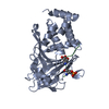 4qumC  2b49S S: Starting model for refinement C: citing same article ( |
|---|---|
| Similar structure data |


-Links
 PDBj
PDBj


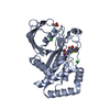
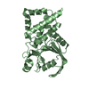
- Assembly
| Deposited unit | 
| ||||||||
|---|---|---|---|---|---|---|---|---|---|
| 1 | 
| ||||||||
| 2 | 
| ||||||||
| Unit cell |
|
-Components
| #1: Protein | Mass: 34931.672 Da / Num. of mol.: 2 / Fragment: Catalytic domain (UNP RESIDUES 628-909) / Mutation: C842S Source method: isolated from a genetically manipulated source Source: (gene. exp.) Homo sapiens (human) / Gene: PTPH1, PTPN3 / Plasmid: pMCSG7 / Production host:  #2: Chemical |   Mass: 94.971 Da / Num. of mol.: 2 / Source method: obtained synthetically / Formula: PO4 Mass: 94.971 Da / Num. of mol.: 2 / Source method: obtained synthetically / Formula: PO4#3: Chemical | ChemComp-GOL / |   Mass: 92.094 Da / Num. of mol.: 1 / Source method: obtained synthetically / Formula: C3H8O3 Mass: 92.094 Da / Num. of mol.: 1 / Source method: obtained synthetically / Formula: C3H8O3#4: Water | ChemComp-HOH / |  Mass: 18.015 Da / Num. of mol.: 338 / Source method: isolated from a natural source / Formula: H2O Mass: 18.015 Da / Num. of mol.: 338 / Source method: isolated from a natural source / Formula: H2O |
|---|
-Experimental details
-Experiment
| Experiment | Method: X-RAY DIFFRACTION / Number of used crystals: 1 |
|---|
- Sample preparation
| Crystal | Density Matthews: 2.27 Å3/Da / Density % sol: 45.87 % |
|---|---|
| Crystal grow | Temperature: 293 K / Method: vapor diffusion, sitting drop / pH: 7.5 Details: 0.1M Hepes, 12% PEG 8000, pH 7.5, VAPOR DIFFUSION, SITTING DROP, temperature 293K |
-Data collection
| Diffraction | Mean temperature: 100 K |
|---|---|
| Diffraction source | Source: ROTATING ANODE / Type: RIGAKU FR-E+ SUPERBRIGHT / Wavelength: 1.54 Å |
| Detector | Type: RIGAKU RAXIS HTC / Detector: IMAGE PLATE / Date: Sep 9, 2012 |
| Radiation | Monochromator: Ni MIRROR + Ni FILTER / Protocol: SINGLE WAVELENGTH / Monochromatic (M) / Laue (L): M / Scattering type: x-ray |
| Radiation wavelength | Wavelength: 1.54 Å / Relative weight: 1 |
| Reflection | Resolution: 1.86→50 Å / Num. all: 53391 / Num. obs: 53391 / % possible obs: 99.5 % / Observed criterion σ(F): 0 / Observed criterion σ(I): 0 / Redundancy: 6.8 % / Biso Wilson estimate: 31.9 Å2 / Rmerge(I) obs: 0.08 / Net I/σ(I): 18.2 |
| Reflection shell | Resolution: 1.86→1.93 Å / Redundancy: 6.8 % / Rmerge(I) obs: 0.88 / Mean I/σ(I) obs: 2.2 / Num. unique all: 5319 / % possible all: 100 |
- Processing
| Software |
| ||||||||||||||||||||||||||||||||||||||||||||||||||||||||||||||||||||||||||||||||||||||||||||||||||||||||||||||||||||||||
|---|---|---|---|---|---|---|---|---|---|---|---|---|---|---|---|---|---|---|---|---|---|---|---|---|---|---|---|---|---|---|---|---|---|---|---|---|---|---|---|---|---|---|---|---|---|---|---|---|---|---|---|---|---|---|---|---|---|---|---|---|---|---|---|---|---|---|---|---|---|---|---|---|---|---|---|---|---|---|---|---|---|---|---|---|---|---|---|---|---|---|---|---|---|---|---|---|---|---|---|---|---|---|---|---|---|---|---|---|---|---|---|---|---|---|---|---|---|---|---|---|---|
| Refinement | Method to determine structure: MOLECULAR REPLACEMENT Starting model: PDB Entry 2B49 Resolution: 1.86→45.764 Å / SU ML: 0.19 / σ(F): 1.34 / Phase error: 19.92 / Stereochemistry target values: MLHL
| ||||||||||||||||||||||||||||||||||||||||||||||||||||||||||||||||||||||||||||||||||||||||||||||||||||||||||||||||||||||||
| Solvent computation | Shrinkage radii: 0.9 Å / VDW probe radii: 1.11 Å / Solvent model: FLAT BULK SOLVENT MODEL | ||||||||||||||||||||||||||||||||||||||||||||||||||||||||||||||||||||||||||||||||||||||||||||||||||||||||||||||||||||||||
| Refinement step | Cycle: LAST / Resolution: 1.86→45.764 Å
| ||||||||||||||||||||||||||||||||||||||||||||||||||||||||||||||||||||||||||||||||||||||||||||||||||||||||||||||||||||||||
| Refine LS restraints |
| ||||||||||||||||||||||||||||||||||||||||||||||||||||||||||||||||||||||||||||||||||||||||||||||||||||||||||||||||||||||||
| LS refinement shell | Refine-ID: X-RAY DIFFRACTION / Total num. of bins used: 19
|
