 Movie
Movie Controller


+ Open data

- Basic information
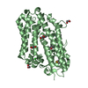
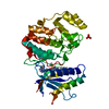
| Entry | Database: PDB / ID: 1cm8 | ||||||
|---|---|---|---|---|---|---|---|
| Title | PHOSPHORYLATED MAP KINASE P38-GAMMA | ||||||
 Components Components | PHOSPHORYLATED MAP KINASE P38-GAMMA | ||||||
 Keywords Keywords | TRANSFERASE / P38-GAMMA / GAMMA / PHOSPHORYLATION / MAP KINASE | ||||||
| Function / homology |  Function and homology information Function and homology informationmyoblast differentiation / Activation of PPARGC1A (PGC-1alpha) by phosphorylation / peptidase activator activity / muscle organ development / positive regulation of muscle cell differentiation / Myogenesis / negative regulation of cell cycle / MAP kinase activity / mitogen-activated protein kinase / signal transduction in response to DNA damage ...myoblast differentiation / Activation of PPARGC1A (PGC-1alpha) by phosphorylation / peptidase activator activity / muscle organ development / positive regulation of muscle cell differentiation / Myogenesis / negative regulation of cell cycle / MAP kinase activity / mitogen-activated protein kinase / signal transduction in response to DNA damage / p38MAPK events / NOD1/2 Signaling Pathway / VEGFA-VEGFR2 Pathway / Negative regulation of MAPK pathway / MAPK cascade / regulation of cell cycle / intracellular signal transduction / protein serine kinase activity / protein serine/threonine kinase activity / magnesium ion binding / signal transduction / mitochondrion / nucleoplasm / ATP binding / nucleus / cytoplasm / cytosol Similarity search - Function | ||||||
| Biological species |  Homo sapiens (human) Homo sapiens (human) | ||||||
| Method |  X-RAY DIFFRACTION / SYNCHROTRON / MIR / Resolution: 2.4 Å X-RAY DIFFRACTION / SYNCHROTRON / MIR / Resolution: 2.4 Å | ||||||
 Authors Authors | Bellon, S. / Fitzgibbon, M.J. / Fox, T. / Hsiao, H.M. / Wilson, K.P. | ||||||
 Citation Citation | Journal: Structure Fold.Des. / Year: 1999 Title: The structure of phosphorylated p38gamma is monomeric and reveals a conserved activation-loop conformation. Authors: Bellon, S. / Fitzgibbon, M.J. / Fox, T. / Hsiao, H.M. / Wilson, K.P. | ||||||
| History |
|
- Structure visualization
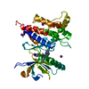
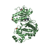
| Structure viewer | Molecule: MolmilJmol/JSmol |
|---|
- Downloads & links
-Download
| PDBx/mmCIF format | 1cm8.cif.gz | 150.7 KB | Display | PDBx/mmCIF format |
|---|---|---|---|---|
| PDB format | pdb1cm8.ent.gz | 115.8 KB | Display | PDB format |
| PDBx/mmJSON format | 1cm8.json.gz | Tree view | PDBx/mmJSON format | |
| Others |  Other downloads Other downloads |
-Validation report
| Arichive directory | https://data.pdbj.org/pub/pdb/validation_reports/cm/1cm8ftp://data.pdbj.org/pub/pdb/validation_reports/cm/1cm8 | HTTPS FTP |
|---|
-Related structure data
| Similar structure data |
|---|


-Links
 PDBj
PDBj


- Assembly
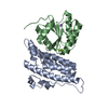
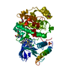
| Deposited unit | 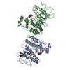
| ||||||||||
|---|---|---|---|---|---|---|---|---|---|---|---|
| 1 | 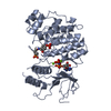
| ||||||||||
| 2 | 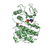
| ||||||||||
| Unit cell |
| ||||||||||
| Noncrystallographic symmetry (NCS) | NCS oper: (Code: given Matrix: (-0.0105, -0.9989, -0.0454), Vector: |
-Components
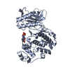
| #1: Protein | Mass: 42157.090 Da / Num. of mol.: 2 Source method: isolated from a genetically manipulated source Source: (gene. exp.) Homo sapiens (human) / Production host:  #2: Chemical | ChemComp-MG /   Mass: 24.305 Da / Num. of mol.: 4 / Source method: obtained synthetically / Formula: Mg Mass: 24.305 Da / Num. of mol.: 4 / Source method: obtained synthetically / Formula: Mg#3: Chemical |   Mass: 506.196 Da / Num. of mol.: 2 / Source method: obtained synthetically / Formula: C10H17N6O12P3 / Comment: AMP-PNP, energy-carrying molecule analogue*YM Mass: 506.196 Da / Num. of mol.: 2 / Source method: obtained synthetically / Formula: C10H17N6O12P3 / Comment: AMP-PNP, energy-carrying molecule analogue*YM#4: Water | ChemComp-HOH / |  Mass: 18.015 Da / Num. of mol.: 186 / Source method: isolated from a natural source / Formula: H2O Mass: 18.015 Da / Num. of mol.: 186 / Source method: isolated from a natural source / Formula: H2OHas protein modification | Y | |
|---|
-Experimental details
-Experiment
| Experiment | Method: X-RAY DIFFRACTION / Number of used crystals: 1 |
|---|
- Sample preparation
| Crystal | Density Matthews: 2.59 Å3/Da / Density % sol: 52.52 % | |||||||||||||||||||||||||||||||||||||||||||||||||||||||||||||||
|---|---|---|---|---|---|---|---|---|---|---|---|---|---|---|---|---|---|---|---|---|---|---|---|---|---|---|---|---|---|---|---|---|---|---|---|---|---|---|---|---|---|---|---|---|---|---|---|---|---|---|---|---|---|---|---|---|---|---|---|---|---|---|---|---|
| Crystal grow | Method: vapor diffusion / pH: 7 / Details: pH 7.0, VAPOR DIFFUSION | |||||||||||||||||||||||||||||||||||||||||||||||||||||||||||||||
| Crystal grow | *PLUS pH: 8.5 | |||||||||||||||||||||||||||||||||||||||||||||||||||||||||||||||
| Components of the solutions | *PLUS
|
-Data collection
| Diffraction | Mean temperature: 110 K |
|---|---|
| Diffraction source | Source: SYNCHROTRON / Site: NSLS  / Beamline: X25 / Wavelength: 1.1 / Beamline: X25 / Wavelength: 1.1 |
| Detector | Type: MARRESEARCH / Detector: IMAGE PLATE |
| Radiation | Protocol: SINGLE WAVELENGTH / Monochromatic (M) / Laue (L): M / Scattering type: x-ray |
| Radiation wavelength | Wavelength: 1.1 Å / Relative weight: 1 |
| Reflection | Resolution: 2.4→50 Å / Num. obs: 31732 / % possible obs: 89.9 % / Redundancy: 3.7 % / Rsym value: 6.7 / Net I/σ(I): 9.8 |
| Reflection | *PLUS Num. measured all: 118429 / Rmerge(I) obs: 0.067 |
| Reflection shell | *PLUS Highest resolution: 2.4 Å / Lowest resolution: 2.49 Å / % possible obs: 76.5 % |
- Processing
| Software |
| ||||||||||||||||||||||||||||||||||||||||||||||||||||||||||||
|---|---|---|---|---|---|---|---|---|---|---|---|---|---|---|---|---|---|---|---|---|---|---|---|---|---|---|---|---|---|---|---|---|---|---|---|---|---|---|---|---|---|---|---|---|---|---|---|---|---|---|---|---|---|---|---|---|---|---|---|---|---|
| Refinement | Method to determine structure: MIR / Resolution: 2.4→50 Å / Rfactor Rfree error: 0.006 / Cross valid method: THROUGHOUT / σ(F): 2 Details: THE DATA ARE EXTREMELY ANISOTROPIC. THIS RESULTED IN LESS THAN 100% COMPLETENESS FOR THE NATIVE DATA.
| ||||||||||||||||||||||||||||||||||||||||||||||||||||||||||||
| Refinement step | Cycle: LAST / Resolution: 2.4→50 Å
| ||||||||||||||||||||||||||||||||||||||||||||||||||||||||||||
| Refine LS restraints |
| ||||||||||||||||||||||||||||||||||||||||||||||||||||||||||||
| Refine LS restraints NCS | NCS model details: RESTRAINTS | ||||||||||||||||||||||||||||||||||||||||||||||||||||||||||||
| Software | *PLUS Name: CNS / Version: 0.2 / Classification: refinement | ||||||||||||||||||||||||||||||||||||||||||||||||||||||||||||
| Refinement | *PLUS Highest resolution: 2.4 Å / Lowest resolution: 50 Å / σ(F): 2 / % reflection Rfree: 9.9 % / Rfactor obs: 0.232 | ||||||||||||||||||||||||||||||||||||||||||||||||||||||||||||
| Solvent computation | *PLUS | ||||||||||||||||||||||||||||||||||||||||||||||||||||||||||||
| Displacement parameters | *PLUS | ||||||||||||||||||||||||||||||||||||||||||||||||||||||||||||
| Refine LS restraints | *PLUS
|
