 Movie
Movie Controller

[English] 日本語
 Yorodumi
- PDB-6w1k: Crystal structure of the hydroxyglutarate synthase in complex wit... -
+ Open data

- Basic information
| Entry | Database: PDB / ID: 6w1k | ||||||
|---|---|---|---|---|---|---|---|
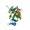
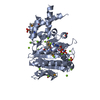
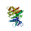
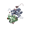
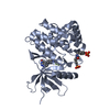
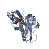
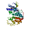
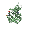
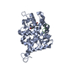
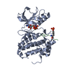
| Title | Crystal structure of the hydroxyglutarate synthase in complex with 2-oxoadipate from Oryza sativa | ||||||
 Components Components | Hydroxyglutarate synthase | ||||||
 Keywords Keywords | LYASE / Decarboxylase Lysine catabolism | ||||||
| Function / homology |  Function and homology information Function and homology information2-oxoadipate dioxygenase/decarboxylase / amyloplast / dioxygenase activity / chloroplast / metal ion binding Similarity search - Function | ||||||
| Biological species |  | ||||||
| Method |  X-RAY DIFFRACTION / SYNCHROTRON / MOLECULAR REPLACEMENT / Resolution: 1.85 Å X-RAY DIFFRACTION / SYNCHROTRON / MOLECULAR REPLACEMENT / Resolution: 1.85 Å | ||||||
 Authors Authors | Pereira, J.H. / Thompson, M.G. / Blake-Hedges, J.M. / Keasling, J.D. / Adams, P.D. | ||||||
 Citation Citation | Journal: Nat Commun / Year: 2020 Title: An iron (II) dependent oxygenase performs the last missing step of plant lysine catabolism. Authors: Thompson, M.G. / Blake-Hedges, J.M. / Pereira, J.H. / Hangasky, J.A. / Belcher, M.S. / Moore, W.M. / Barajas, J.F. / Cruz-Morales, P. / Washington, L.J. / Haushalter, R.W. / Eiben, C.B. / ...Authors: Thompson, M.G. / Blake-Hedges, J.M. / Pereira, J.H. / Hangasky, J.A. / Belcher, M.S. / Moore, W.M. / Barajas, J.F. / Cruz-Morales, P. / Washington, L.J. / Haushalter, R.W. / Eiben, C.B. / Liu, Y. / Skyrud, W. / Benites, V.T. / Barnum, T.P. / Baidoo, E.E.K. / Scheller, H.V. / Marletta, M.A. / Shih, P.M. / Adams, P.D. / Keasling, J.D. | ||||||
| History |
|
- Structure visualization
| Structure viewer | Molecule: MolmilJmol/JSmol |
|---|
- Downloads & links
-Download
| PDBx/mmCIF format | 6w1k.cif.gz | 763.6 KB | Display | PDBx/mmCIF format |
|---|---|---|---|---|
| PDB format | pdb6w1k.ent.gz | 620 KB | Display | PDB format |
| PDBx/mmJSON format | 6w1k.json.gz | Tree view | PDBx/mmJSON format | |
| Others |  Other downloads Other downloads |
-Validation report
| Arichive directory | https://data.pdbj.org/pub/pdb/validation_reports/w1/6w1kftp://data.pdbj.org/pub/pdb/validation_reports/w1/6w1k | HTTPS FTP |
|---|
-Related structure data
| Related structure data | 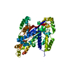 6w1gC 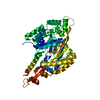 6w1hC  3lhoS S: Starting model for refinement C: citing same article ( |
|---|---|
| Similar structure data |
-Links
 PDBj
PDBj- Assembly
| Deposited unit | 
| ||||||||||||
|---|---|---|---|---|---|---|---|---|---|---|---|---|---|
| 1 | 
| ||||||||||||
| 2 | 
| ||||||||||||
| 3 | 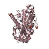
| ||||||||||||
| 4 | 
| ||||||||||||
| Unit cell |
|
-Components
| #1: Protein | Mass: 36140.234 Da / Num. of mol.: 4 Source method: isolated from a genetically manipulated source Source: (gene. exp.)  #2: Chemical | ChemComp-NI /   Mass: 58.693 Da / Num. of mol.: 4 / Source method: obtained synthetically / Formula: Ni Mass: 58.693 Da / Num. of mol.: 4 / Source method: obtained synthetically / Formula: Ni#3: Chemical | ChemComp-OOG /   Mass: 160.125 Da / Num. of mol.: 4 / Source method: obtained synthetically / Formula: C6H8O5 / Feature type: SUBJECT OF INVESTIGATION Mass: 160.125 Da / Num. of mol.: 4 / Source method: obtained synthetically / Formula: C6H8O5 / Feature type: SUBJECT OF INVESTIGATION#4: Chemical | ChemComp-SO4 /   Mass: 96.063 Da / Num. of mol.: 16 / Source method: obtained synthetically / Formula: SO4 Mass: 96.063 Da / Num. of mol.: 16 / Source method: obtained synthetically / Formula: SO4#5: Water | ChemComp-HOH / |  Mass: 18.015 Da / Num. of mol.: 1123 / Source method: isolated from a natural source / Formula: H2O Mass: 18.015 Da / Num. of mol.: 1123 / Source method: isolated from a natural source / Formula: H2OHas ligand of interest | Y | |
|---|
-Experimental details
-Experiment
| Experiment | Method: X-RAY DIFFRACTION / Number of used crystals: 1 |
|---|
- Sample preparation
| Crystal | Density Matthews: 3.19 Å3/Da / Density % sol: 61.5 % |
|---|---|
| Crystal grow | Temperature: 298 K / Method: vapor diffusion, sitting drop / pH: 5.6 Details: 0.01 M Magnesium Chloride, 0.05 M MES pH 5.6, 1.8 M Lithium Sulfate |
-Data collection
| Diffraction | Mean temperature: 100 K / Serial crystal experiment: N |
|---|---|
| Diffraction source | Source: SYNCHROTRON / Site: SSRL  / Beamline: BL12-2 / Wavelength: 0.97946 Å / Beamline: BL12-2 / Wavelength: 0.97946 Å |
| Detector | Type: DECTRIS PILATUS 6M / Detector: PIXEL / Date: Feb 19, 2019 |
| Radiation | Protocol: SINGLE WAVELENGTH / Monochromatic (M) / Laue (L): M / Scattering type: x-ray |
| Radiation wavelength | Wavelength: 0.97946 Å / Relative weight: 1 |
| Reflection | Resolution: 1.85→39.8 Å / Num. obs: 156493 / % possible obs: 98.9 % / Redundancy: 7.3 % / Biso Wilson estimate: 29.19 Å2 / CC1/2: 0.998 / Net I/σ(I): 11.5 |
| Reflection shell | Resolution: 1.85→1.91 Å / Num. unique obs: 15364 / CC1/2: 0.517 |
- Processing
| Software |
| |||||||||||||||||||||||||||||||||||||||||||||||||||||||||||||||||||||||||||||||||||||||||||||||||||||||||||||||||||||||||||||||||||||||||||||||||||||||||||||||||||||||||||||||||||||||||||||||||||||||||||||||||||||||||
|---|---|---|---|---|---|---|---|---|---|---|---|---|---|---|---|---|---|---|---|---|---|---|---|---|---|---|---|---|---|---|---|---|---|---|---|---|---|---|---|---|---|---|---|---|---|---|---|---|---|---|---|---|---|---|---|---|---|---|---|---|---|---|---|---|---|---|---|---|---|---|---|---|---|---|---|---|---|---|---|---|---|---|---|---|---|---|---|---|---|---|---|---|---|---|---|---|---|---|---|---|---|---|---|---|---|---|---|---|---|---|---|---|---|---|---|---|---|---|---|---|---|---|---|---|---|---|---|---|---|---|---|---|---|---|---|---|---|---|---|---|---|---|---|---|---|---|---|---|---|---|---|---|---|---|---|---|---|---|---|---|---|---|---|---|---|---|---|---|---|---|---|---|---|---|---|---|---|---|---|---|---|---|---|---|---|---|---|---|---|---|---|---|---|---|---|---|---|---|---|---|---|---|---|---|---|---|---|---|---|---|---|---|---|---|---|---|---|---|
| Refinement | Method to determine structure: MOLECULAR REPLACEMENT Starting model: 3LHO Resolution: 1.85→39.79 Å / SU ML: 0.189 / Cross valid method: FREE R-VALUE / σ(F): 1.34 / Phase error: 19.4898
| |||||||||||||||||||||||||||||||||||||||||||||||||||||||||||||||||||||||||||||||||||||||||||||||||||||||||||||||||||||||||||||||||||||||||||||||||||||||||||||||||||||||||||||||||||||||||||||||||||||||||||||||||||||||||
| Solvent computation | Shrinkage radii: 0.9 Å / VDW probe radii: 1.11 Å | |||||||||||||||||||||||||||||||||||||||||||||||||||||||||||||||||||||||||||||||||||||||||||||||||||||||||||||||||||||||||||||||||||||||||||||||||||||||||||||||||||||||||||||||||||||||||||||||||||||||||||||||||||||||||
| Displacement parameters | Biso mean: 39.28 Å2 | |||||||||||||||||||||||||||||||||||||||||||||||||||||||||||||||||||||||||||||||||||||||||||||||||||||||||||||||||||||||||||||||||||||||||||||||||||||||||||||||||||||||||||||||||||||||||||||||||||||||||||||||||||||||||
| Refinement step | Cycle: LAST / Resolution: 1.85→39.79 Å
| |||||||||||||||||||||||||||||||||||||||||||||||||||||||||||||||||||||||||||||||||||||||||||||||||||||||||||||||||||||||||||||||||||||||||||||||||||||||||||||||||||||||||||||||||||||||||||||||||||||||||||||||||||||||||
| Refine LS restraints |
| |||||||||||||||||||||||||||||||||||||||||||||||||||||||||||||||||||||||||||||||||||||||||||||||||||||||||||||||||||||||||||||||||||||||||||||||||||||||||||||||||||||||||||||||||||||||||||||||||||||||||||||||||||||||||
| LS refinement shell |
|
