 Movie
Movie Controller

[English] 日本語
 Yorodumi
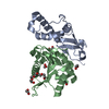
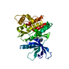
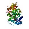
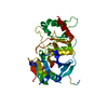
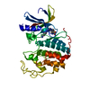
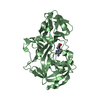
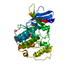
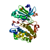
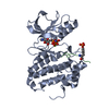
- PDB-6b92: Crystal Structure of the N-terminal domain of human METTL16 in co... -
+ Open data

- Basic information
| Entry | Database: PDB / ID: 6b92 | ||||||
|---|---|---|---|---|---|---|---|
| Title | Crystal Structure of the N-terminal domain of human METTL16 in complex with SAH | ||||||
 Components Components | U6 small nuclear RNA (adenine-(43)-N(6))-methyltransferase | ||||||
 Keywords Keywords | TRANSFERASE / methyltransferase-like protein 16 / RNA methylation / N6-methyladenosine / S-adenosylmethionine / S-adenosylhomocysteine | ||||||
| Function / homology |  Function and homology information Function and homology informationsnRNA (adenine-N6)-methylation / U6 snRNA m6A methyltransferase / U6 snRNA (adenine(43)-N6)-methyltransferase activity / 23S rRNA (adenine(1618)-N(6))-methyltransferase activity / negative regulation of 3'-UTR-mediated mRNA stabilization / U6 snRNA 3'-end binding / mRNA m(6)A methyltransferase activity / mRNA m6A methyltransferase / S-adenosylmethionine biosynthetic process / regulation of mRNA splicing, via spliceosome ...snRNA (adenine-N6)-methylation / U6 snRNA m6A methyltransferase / U6 snRNA (adenine(43)-N6)-methyltransferase activity / 23S rRNA (adenine(1618)-N(6))-methyltransferase activity / negative regulation of 3'-UTR-mediated mRNA stabilization / U6 snRNA 3'-end binding / mRNA m(6)A methyltransferase activity / mRNA m6A methyltransferase / S-adenosylmethionine biosynthetic process / regulation of mRNA splicing, via spliceosome / rRNA base methylation / post-transcriptional regulation of gene expression / mRNA destabilization / mRNA catabolic process / RNA stem-loop binding / mRNA processing / RNA binding / nucleus / cytoplasm Similarity search - Function | ||||||
| Biological species |  Homo sapiens (human) Homo sapiens (human) | ||||||
| Method |  X-RAY DIFFRACTION / SYNCHROTRON / MOLECULAR REPLACEMENT / Resolution: 2.1 Å X-RAY DIFFRACTION / SYNCHROTRON / MOLECULAR REPLACEMENT / Resolution: 2.1 Å | ||||||
 Authors Authors | Ruszkowska, A. / Ruszkowski, M. / Dauter, Z. / Brown, J.A. | ||||||
| Funding support |  United States, 1items United States, 1items
| ||||||
 Citation Citation | Journal: Sci Rep / Year: 2018 Title: Structural insights into the RNA methyltransferase domain of METTL16. Authors: Ruszkowska, A. / Ruszkowski, M. / Dauter, Z. / Brown, J.A. | ||||||
| History |
|
- Structure visualization
| Structure viewer | Molecule: MolmilJmol/JSmol |
|---|
- Downloads & links
-Download
| PDBx/mmCIF format | 6b92.cif.gz | 130.2 KB | Display | PDBx/mmCIF format |
|---|---|---|---|---|
| PDB format | pdb6b92.ent.gz | 100.2 KB | Display | PDB format |
| PDBx/mmJSON format | 6b92.json.gz | Tree view | PDBx/mmJSON format | |
| Others |  Other downloads Other downloads |
-Validation report
| Arichive directory | https://data.pdbj.org/pub/pdb/validation_reports/b9/6b92ftp://data.pdbj.org/pub/pdb/validation_reports/b9/6b92 | HTTPS FTP |
|---|
-Related structure data
| Related structure data | 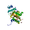 6b91C  2h00S C: citing same article ( S: Starting model for refinement |
|---|---|
| Similar structure data |

-Links
 PDBj
PDBj- Assembly
| Deposited unit | 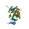
| |||||||||
|---|---|---|---|---|---|---|---|---|---|---|
| 1 |
| |||||||||
| Unit cell |
| |||||||||
| Components on special symmetry positions |
|
-Components
| #1: Protein | Mass: 33617.797 Da / Num. of mol.: 1 / Fragment: N-terminal domain Source method: isolated from a genetically manipulated source Source: (gene. exp.) Homo sapiens (human) / Gene: METTL16, METT10DProduction host:  References: UniProt: Q86W50, Transferases; Transferring one-carbon groups; Methyltransferases, mRNA (2'-O-methyladenosine-N6-)-methyltransferase | ||||
|---|---|---|---|---|---|
| #2: Chemical | ChemComp-EDO /   Mass: 62.068 Da / Num. of mol.: 4 / Source method: obtained synthetically / Formula: C2H6O2 Mass: 62.068 Da / Num. of mol.: 4 / Source method: obtained synthetically / Formula: C2H6O2#3: Chemical | ChemComp-SAH / |   Type: L-peptide linking / Mass: 384.411 Da / Num. of mol.: 1 / Source method: obtained synthetically / Formula: C14H20N6O5S Type: L-peptide linking / Mass: 384.411 Da / Num. of mol.: 1 / Source method: obtained synthetically / Formula: C14H20N6O5S#4: Water | ChemComp-HOH / |  Mass: 18.015 Da / Num. of mol.: 181 / Source method: isolated from a natural source / Formula: H2O Mass: 18.015 Da / Num. of mol.: 181 / Source method: isolated from a natural source / Formula: H2O |
-Experimental details
-Experiment
| Experiment | Method: X-RAY DIFFRACTION / Number of used crystals: 1 |
|---|
- Sample preparation
| Crystal | Density Matthews: 4.77 Å3/Da / Density % sol: 74.23 % |
|---|---|
| Crystal grow | Temperature: 293 K / Method: vapor diffusion, hanging drop / pH: 8.5 Details: 1.3 M K2HPO4, 45 mM NaH2PO4; drop contained 4 uL of protein solution (27 mg/mL) and 2 uL of reservoir solution; 0.2 uL of 200 mM SAM solution (buffered in 50 mM Hepes pH7.5) was added to the ...Details: 1.3 M K2HPO4, 45 mM NaH2PO4; drop contained 4 uL of protein solution (27 mg/mL) and 2 uL of reservoir solution; 0.2 uL of 200 mM SAM solution (buffered in 50 mM Hepes pH7.5) was added to the drop with mature crystals. 25% ethylene glycol was used for cryo-protection |
-Data collection
| Diffraction | Mean temperature: 100 K |
|---|---|
| Diffraction source | Source: SYNCHROTRON / Site: APS / Beamline: 22-ID / Wavelength: 1 Å |
| Detector | Type: RAYONIX MX300-HS / Detector: CCD / Date: Jun 25, 2017 |
| Radiation | Monochromator: Si (111) / Protocol: SINGLE WAVELENGTH / Monochromatic (M) / Laue (L): M / Scattering type: x-ray |
| Radiation wavelength | Wavelength: 1 Å / Relative weight: 1 |
| Reflection | Resolution: 2.1→50 Å / Num. obs: 34224 / % possible obs: 99.3 % / Redundancy: 17.3 % / CC1/2: 1 / Rmerge(I) obs: 0.064 / Rrim(I) all: 0.066 / Net I/σ(I): 28.7 |
| Reflection shell | Resolution: 2.1→2.23 Å / Redundancy: 14.2 % / Rmerge(I) obs: 1.36 / Mean I/σ(I) obs: 2 / Num. unique obs: 5267 / CC1/2: 0.72 / Rrim(I) all: 1.41 / % possible all: 95.9 |
- Processing
| Software |
| ||||||||||||||||||||||||||||||||||||||||||||||||||||||||||||||||||||||||||||||||||||||||||||||||||||||||||||||||||||||||||||||||||||||||||||||||||||||||||||||||||||||||||||||||||||||
|---|---|---|---|---|---|---|---|---|---|---|---|---|---|---|---|---|---|---|---|---|---|---|---|---|---|---|---|---|---|---|---|---|---|---|---|---|---|---|---|---|---|---|---|---|---|---|---|---|---|---|---|---|---|---|---|---|---|---|---|---|---|---|---|---|---|---|---|---|---|---|---|---|---|---|---|---|---|---|---|---|---|---|---|---|---|---|---|---|---|---|---|---|---|---|---|---|---|---|---|---|---|---|---|---|---|---|---|---|---|---|---|---|---|---|---|---|---|---|---|---|---|---|---|---|---|---|---|---|---|---|---|---|---|---|---|---|---|---|---|---|---|---|---|---|---|---|---|---|---|---|---|---|---|---|---|---|---|---|---|---|---|---|---|---|---|---|---|---|---|---|---|---|---|---|---|---|---|---|---|---|---|---|---|
| Refinement | Method to determine structure: MOLECULAR REPLACEMENT Starting model: 2h00 Resolution: 2.1→44.87 Å / Cor.coef. Fo:Fc: 0.968 / Cor.coef. Fo:Fc free: 0.953 / SU B: 7.465 / SU ML: 0.098 / Cross valid method: FREE R-VALUE / ESU R: 0.122 / ESU R Free: 0.125 Details: HYDROGENS WERE ADDED IN THE RIDING POSITIONS FOR REFINEMENT
| ||||||||||||||||||||||||||||||||||||||||||||||||||||||||||||||||||||||||||||||||||||||||||||||||||||||||||||||||||||||||||||||||||||||||||||||||||||||||||||||||||||||||||||||||||||||
| Solvent computation | Ion probe radii: 0.8 Å / Shrinkage radii: 0.8 Å / VDW probe radii: 1.2 Å | ||||||||||||||||||||||||||||||||||||||||||||||||||||||||||||||||||||||||||||||||||||||||||||||||||||||||||||||||||||||||||||||||||||||||||||||||||||||||||||||||||||||||||||||||||||||
| Displacement parameters | Biso mean: 54.009 Å2
| ||||||||||||||||||||||||||||||||||||||||||||||||||||||||||||||||||||||||||||||||||||||||||||||||||||||||||||||||||||||||||||||||||||||||||||||||||||||||||||||||||||||||||||||||||||||
| Refinement step | Cycle: 1 / Resolution: 2.1→44.87 Å
| ||||||||||||||||||||||||||||||||||||||||||||||||||||||||||||||||||||||||||||||||||||||||||||||||||||||||||||||||||||||||||||||||||||||||||||||||||||||||||||||||||||||||||||||||||||||
| Refine LS restraints |
|