 Movie
Movie Controller


+ Open data

- Basic information
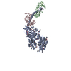
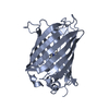
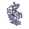
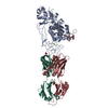
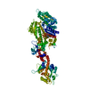
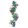
| Entry | Database: PDB / ID: 4p7h | |||||||||
|---|---|---|---|---|---|---|---|---|---|---|
| Title | Structure of Human beta-Cardiac Myosin Motor Domain::GFP chimera | |||||||||
 Components Components | Myosin-7,Green fluorescent protein | |||||||||
 Keywords Keywords | Motor/Fluorescent Protein / Cardiac / Motor / Motor-Fluorescent Protein Complex | |||||||||
| Function / homology |  Function and homology information Function and homology informationregulation of slow-twitch skeletal muscle fiber contraction / regulation of the force of skeletal muscle contraction / transition between fast and slow fiber / regulation of the force of heart contraction / cardiac muscle hypertrophy in response to stress / muscle myosin complex / adult heart development / myosin filament / muscle filament sliding / myosin complex ...regulation of slow-twitch skeletal muscle fiber contraction / regulation of the force of skeletal muscle contraction / transition between fast and slow fiber / regulation of the force of heart contraction / cardiac muscle hypertrophy in response to stress / muscle myosin complex / adult heart development / myosin filament / muscle filament sliding / myosin complex / myosin II complex / ventricular cardiac muscle tissue morphogenesis / microfilament motor activity / myofibril / ATP metabolic process / cardiac muscle contraction / skeletal muscle contraction / stress fiber / muscle contraction / regulation of heart rate / bioluminescence / striated muscle contraction / sarcomere / generation of precursor metabolites and energy / Z disc / actin filament binding / calmodulin binding / ATP binding / cytoplasm Similarity search - Function | |||||||||
| Biological species |  Homo sapiens (human) Homo sapiens (human)  Aequorea victoria (jellyfish) Aequorea victoria (jellyfish) | |||||||||
| Method |  X-RAY DIFFRACTION / MOLECULAR REPLACEMENT / Resolution: 3.2 Å X-RAY DIFFRACTION / MOLECULAR REPLACEMENT / Resolution: 3.2 Å | |||||||||
 Authors Authors | Winkelmann, D.A. / Miller, M.T. / Stock, A.M. | |||||||||
| Funding support |  United States, 1items United States, 1items
| |||||||||
 Citation Citation | Journal: Mol. Biol. Cell / Year: 2011 Title: Structure of Human beta-Cardiac Myosin Motor Domain at 3.2 A Authors: Winkelmann, D.A. / Miller, M.T. / Stock, A.M. / Liu, L. #1: Journal: J.Biol.Chem. / Year: 2002 Title: Folding of the striated muscle myosin motor domain. Authors: Chow, D. / Srikakulam, R. / Chen, Y. / Winkelmann, D.A. | |||||||||
| History |
|
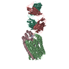
- Structure visualization
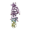
| Structure viewer | Molecule: MolmilJmol/JSmol |
|---|
- Downloads & links
-Download
| PDBx/mmCIF format | 4p7h.cif.gz | 392.7 KB | Display | PDBx/mmCIF format |
|---|---|---|---|---|
| PDB format | pdb4p7h.ent.gz | 314.4 KB | Display | PDB format |
| PDBx/mmJSON format | 4p7h.json.gz | Tree view | PDBx/mmJSON format | |
| Others |  Other downloads Other downloads |
-Validation report
| Arichive directory | https://data.pdbj.org/pub/pdb/validation_reports/p7/4p7hftp://data.pdbj.org/pub/pdb/validation_reports/p7/4p7h | HTTPS FTP |
|---|
-Related structure data


-Links
 PDBj
PDBj


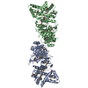
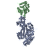
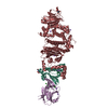
- Assembly
| Deposited unit | 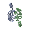
| ||||||||
|---|---|---|---|---|---|---|---|---|---|
| 1 | 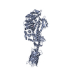
| ||||||||
| 2 | 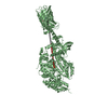
| ||||||||
| Unit cell |
|
-Components
| #1: Protein | Mass: 116468.672 Da / Num. of mol.: 2 Fragment: UNP P12883 residues 1-787,UNP P42212 residues 5-238 Mutation: Q80R, K101N, V163A, I167T, S175G, D190N Source method: isolated from a genetically manipulated source Source: (gene. exp.) Homo sapiens (human), (gene. exp.) Aequorea victoria (jellyfish)Tissue: muscle / Gene: MYH7, MYHCB, GFP / Organ: heart / Cell (production host): myoblast / Cell line (production host): C2C12 / Organ (production host): skeletal muscle / Production host:  #2: Chemical |   Mass: 96.063 Da / Num. of mol.: 2 / Source method: obtained synthetically / Formula: SO4 Mass: 96.063 Da / Num. of mol.: 2 / Source method: obtained synthetically / Formula: SO4#3: Water | ChemComp-HOH / |  Mass: 18.015 Da / Num. of mol.: 5 / Source method: isolated from a natural source / Formula: H2O Mass: 18.015 Da / Num. of mol.: 5 / Source method: isolated from a natural source / Formula: H2OHas protein modification | Y | Sequence details | The mutations in GFP were engineered to enhance folding | |
|---|
-Experimental details
-Experiment
| Experiment | Method: X-RAY DIFFRACTION |
|---|
- Sample preparation
| Crystal | Density Matthews: 2.67 Å3/Da / Density % sol: 53.95 % / Description: small orthorhombic crystals with green tint |
|---|---|
| Crystal grow | Temperature: 293 K / Method: vapor diffusion, hanging drop / pH: 6 Details: 10% Tacsimate, pH 6.0, 10% glycerol, 14-15% PEG 3350, 0.2 mM MgCL2, and 5 mM TCEP PH range: 5.8-6.2 |
-Data collection
| Diffraction | Mean temperature: 100 K |
|---|---|
| Diffraction source | Source: ROTATING ANODE / Type: RIGAKU MICROMAX-007 HF / Wavelength: 1.54056 Å |
| Detector | Type: RIGAKU RAXIS IV++ / Detector: IMAGE PLATE / Date: May 25, 2011 / Details: Varimax HR |
| Radiation | Protocol: SINGLE WAVELENGTH / Monochromatic (M) / Laue (L): M / Scattering type: x-ray |
| Radiation wavelength | Wavelength: 1.54056 Å / Relative weight: 1 |
| Reflection | Resolution: 3.2→39.56 Å / Num. obs: 36487 / % possible obs: 90.4 % / Redundancy: 6.7 % / Net I/σ(I): 8.1 |
| Reflection shell | Resolution: 3.2→3.37 Å / Redundancy: 6.6 % / Mean I/σ(I) obs: 1.8 / % possible all: 81.2 |
- Processing
| Software | Name: PHENIX / Version: (phenix.refine: 1.8.4_1496) / Classification: refinement | |||||||||||||||||||||||||||||||||||||||||||||||||||||||||||||||||||||||||||||||||||||||||||||||||||||||||
|---|---|---|---|---|---|---|---|---|---|---|---|---|---|---|---|---|---|---|---|---|---|---|---|---|---|---|---|---|---|---|---|---|---|---|---|---|---|---|---|---|---|---|---|---|---|---|---|---|---|---|---|---|---|---|---|---|---|---|---|---|---|---|---|---|---|---|---|---|---|---|---|---|---|---|---|---|---|---|---|---|---|---|---|---|---|---|---|---|---|---|---|---|---|---|---|---|---|---|---|---|---|---|---|---|---|---|
| Refinement | Method to determine structure: MOLECULAR REPLACEMENT Starting model: PDB entries 2MYS, 2QLE Resolution: 3.2→37.939 Å / SU ML: 0.5 / Cross valid method: FREE R-VALUE / σ(F): 1.96 / Phase error: 30.65 / Stereochemistry target values: ML
| |||||||||||||||||||||||||||||||||||||||||||||||||||||||||||||||||||||||||||||||||||||||||||||||||||||||||
| Solvent computation | Shrinkage radii: 0.8 Å / VDW probe radii: 1.1 Å / Solvent model: FLAT BULK SOLVENT MODEL | |||||||||||||||||||||||||||||||||||||||||||||||||||||||||||||||||||||||||||||||||||||||||||||||||||||||||
| Refinement step | Cycle: LAST / Resolution: 3.2→37.939 Å
| |||||||||||||||||||||||||||||||||||||||||||||||||||||||||||||||||||||||||||||||||||||||||||||||||||||||||
| Refine LS restraints |
| |||||||||||||||||||||||||||||||||||||||||||||||||||||||||||||||||||||||||||||||||||||||||||||||||||||||||
| LS refinement shell |
|
