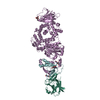
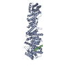
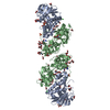
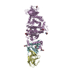
[English] 日本語
Yorodumi
- PDB-7b7n: Human herpesvirus-8 gH/gL in complex with EphA2 -

+
Open data


ID or keywords:

Loading...

-
Basic information

Entry
Database: PDB / ID: 7b7n
TitleHuman herpesvirus-8 gH/gL in complex with EphA2
Components
  • (Envelope glycoprotein ...) x 2
  • Ephrin type-A receptor 2
KeywordsVIRAL PROTEIN / Complex / Receptor
Function / homology
Function and homology information


notochord cell development / notochord formation / lens fiber cell morphogenesis / blood vessel endothelial cell proliferation involved in sprouting angiogenesis / negative regulation of lymphangiogenesis / axial mesoderm formation / pericyte cell differentiation / cAMP metabolic process / virion component => GO:0044423 / positive regulation of bicellular tight junction assembly ...notochord cell development / notochord formation / lens fiber cell morphogenesis / blood vessel endothelial cell proliferation involved in sprouting angiogenesis / negative regulation of lymphangiogenesis / axial mesoderm formation / pericyte cell differentiation / cAMP metabolic process / virion component => GO:0044423 / positive regulation of bicellular tight junction assembly / regulation of blood vessel endothelial cell migration / negative regulation of chemokine production / ephrin receptor activity / leading edge membrane / bone remodeling / post-anal tail morphogenesis / response to growth factor / activation of GTPase activity / regulation of lamellipodium assembly / tight junction / branching involved in mammary gland duct morphogenesis / EPH-Ephrin signaling / neural tube development / RND1 GTPase cycle / RND2 GTPase cycle / RND3 GTPase cycle / mammary gland epithelial cell proliferation / RHOV GTPase cycle / EPHA-mediated growth cone collapse / growth factor binding / regulation of cell adhesion mediated by integrin / lamellipodium membrane / RHOU GTPase cycle / RHOG GTPase cycle / EPH-ephrin mediated repulsion of cells / negative regulation of phosphatidylinositol 3-kinase/protein kinase B signal transduction / RAC3 GTPase cycle / RAC2 GTPase cycle / ephrin receptor signaling pathway / vasculogenesis / regulation of angiogenesis / keratinocyte differentiation / RAC1 GTPase cycle / transmembrane receptor protein tyrosine kinase activity / cell chemotaxis / negative regulation of angiogenesis / osteoclast differentiation / regulation of ERK1 and ERK2 cascade / host cell endosome membrane / phosphatidylinositol 3-kinase/protein kinase B signal transduction / skeletal system development / molecular function activator activity / cell motility / protein localization to plasma membrane / positive regulation of protein localization to plasma membrane / receptor protein-tyrosine kinase / neuron differentiation / ruffle membrane / osteoblast differentiation / cell surface receptor protein tyrosine kinase signaling pathway / intrinsic apoptotic signaling pathway in response to DNA damage / cell migration / virus receptor activity / lamellipodium / host cell Golgi apparatus / receptor complex / cell adhesion / positive regulation of cell migration / defense response to Gram-positive bacterium / cadherin binding / inflammatory response / symbiont entry into host cell / fusion of virus membrane with host plasma membrane / phosphorylation / focal adhesion / viral envelope / host cell plasma membrane / virion membrane / cell surface / ATP binding / membrane / plasma membrane
Similarity search - Function
Herpesvirus glycoprotein L, rhadinovirus-type / Herpesvirus glycoprotein L, rhadinovirus-type superfamily / Viral glycoprotein L / Envelope glycoprotein L / Herpesvirus glycoprotein H main domain / Herpesvirus glycoprotein H / Herpesvirus glycoprotein H, C-terminal / Herpesvirus glycoprotein H, C-terminal domain superfamily / Herpesvirus glycoprotein H C-terminal domain / Ephrin type-A receptor 2, ligand binding domain ...Herpesvirus glycoprotein L, rhadinovirus-type / Herpesvirus glycoprotein L, rhadinovirus-type superfamily / Viral glycoprotein L / Envelope glycoprotein L / Herpesvirus glycoprotein H main domain / Herpesvirus glycoprotein H / Herpesvirus glycoprotein H, C-terminal / Herpesvirus glycoprotein H, C-terminal domain superfamily / Herpesvirus glycoprotein H C-terminal domain / Ephrin type-A receptor 2, ligand binding domain / Ephrin receptor type-A /type-B / Ephrin receptor ligand binding domain / Tyrosine-protein kinase, receptor class V, conserved site / Ephrin receptor, transmembrane domain / Ephrin receptor ligand binding domain / Ephrin type-A receptor 2 transmembrane domain / Receptor tyrosine kinase class V signature 1. / Receptor tyrosine kinase class V signature 2. / Eph receptor ligand-binding domain profile. / Ephrin receptor ligand binding domain / Putative ephrin-receptor like / SAM domain (Sterile alpha motif) / SAM domain profile. / Sterile alpha motif. / Sterile alpha motif domain / Sterile alpha motif/pointed domain superfamily / Growth factor receptor cysteine-rich domain superfamily / Fibronectin type III domain / Fibronectin type 3 domain / Fibronectin type-III domain profile. / Galactose-binding-like domain superfamily / Fibronectin type III / Fibronectin type III superfamily / Tyrosine-protein kinase, catalytic domain / Tyrosine kinase, catalytic domain / Tyrosine protein kinases specific active-site signature. / Tyrosine-protein kinase, active site / Protein tyrosine and serine/threonine kinase / Serine-threonine/tyrosine-protein kinase, catalytic domain / Protein kinase, ATP binding site / Protein kinases ATP-binding region signature. / Immunoglobulin-like fold / Protein kinase domain profile. / Protein kinase domain / Protein kinase-like domain superfamily
Similarity search - Domain/homology
Envelope glycoprotein L / Ephrin type-A receptor 2 / Envelope glycoprotein L / Envelope glycoprotein H
Similarity search - Component
Biological speciesHomo sapiens (human)
Human herpesvirus 8
MethodX-RAY DIFFRACTION / SYNCHROTRON / MOLECULAR REPLACEMENT / Resolution: 2.69 Å
AuthorsPederzoli, R. / Guardado-Calvo, P. / Rey, F.A. / Backovic, M.
Funding support France, 1items
OrganizationGrant numberCountry
Pasteur InstituteRecurrent funding France
CitationJournal: Plos Biol. / Year: 2021
Title: Human herpesvirus 8 molecular mimicry of ephrin ligands facilitates cell entry and triggers EphA2 signaling.
Authors: Light, T.P. / Brun, D. / Guardado-Calvo, P. / Pederzoli, R. / Haouz, A. / Neipel, F. / Rey, F.A. / Hristova, K. / Backovic, M.
History
DepositionDec 11, 2020Deposition site: PDBE / Processing site: PDBE
Revision 1.0Dec 30, 2020Provider: repository / Type: Initial release
Revision 1.1Sep 22, 2021Group: Data collection / Database references
Category: citation / citation_author ...citation / citation_author / database_2 / pdbx_database_proc
Item: _citation.country / _citation.journal_abbrev ..._citation.country / _citation.journal_abbrev / _citation.journal_id_CSD / _citation.journal_id_ISSN / _citation.journal_volume / _citation.page_first / _citation.page_last / _citation.pdbx_database_id_DOI / _citation.pdbx_database_id_PubMed / _citation.title / _citation.year / _database_2.pdbx_DOI / _database_2.pdbx_database_accession
Revision 1.2Jan 31, 2024Group: Data collection / Refinement description
Category: chem_comp_atom / chem_comp_bond / pdbx_initial_refinement_model

-
Structure visualization

Structure viewerMolecule:
MolmilJmol/JSmol

Downloads & links

-
Assembly

Deposited unit
E: Ephrin type-A receptor 2
H: Envelope glycoprotein H
L: Envelope glycoprotein L
hetero molecules


Theoretical massNumber of molelcules
Total (without water)121,37721
Polymers119,7143
Non-polymers1,66318
Water97354
1


  • Idetical with deposited unit
  • defined by author&software
  • Evidence: gel filtration, light scattering
TypeNameSymmetry operationNumber
identity operation1_555x,y,z1
Buried area10310 Å2
ΔGint-164 kcal/mol
Surface area39030 Å2
MethodPISA
Unit cell
Length a, b, c (Å)72.940, 129.000, 267.690
Angle α, β, γ (deg.)90.000, 90.000, 90.000
Int Tables number20
Space group name H-MC2221
Space group name HallC2c2

-
Components

-
Protein , 1 types, 1 molecules E

#1: Protein Ephrin type-A receptor 2 / Epithelial cell kinase / Tyrosine-protein kinase receptor ECK


Mass: 24043.883 Da / Num. of mol.: 1
Source method: isolated from a genetically manipulated source
Source: (gene. exp.) Homo sapiens (human) / Gene: EPHA2, ECK / Cell line (production host): Schneider's Drosophila Line 2 / Production host: Drosophila melanogaster (fruit fly)
References: UniProt: P29317, receptor protein-tyrosine kinase

-
Envelope glycoprotein ... , 2 types, 2 molecules HL

#2: Protein Envelope glycoprotein H / gH


Mass: 76018.961 Da / Num. of mol.: 1
Source method: isolated from a genetically manipulated source
Source: (gene. exp.) Human herpesvirus 8 / Gene: ORF22, gH, HHV8GK18_gp26 / Plasmid: pT350 / Cell line (production host): Schneider's Drosophila Line 2 / Production host: Drosophila melanogaster (fruit fly) / References: UniProt: Q98142
#3: Protein Envelope glycoprotein L / gL


Mass: 19651.018 Da / Num. of mol.: 1 / Mutation: C58S
Source method: isolated from a genetically manipulated source
Source: (gene. exp.) Human herpesvirus 8 / Gene: ORF47, gL, ORF46, HHV8GK18_gp49 / Plasmid: pT350 / Cell line (production host): Schneider's Drosophila Line 2 / Production host: Drosophila melanogaster (fruit fly) / References: UniProt: Q76RG7, UniProt: F5HDB7*PLUS

-
Sugars , 2 types, 4 molecules

#4: Polysaccharide 2-acetamido-2-deoxy-beta-D-glucopyranose-(1-4)-2-acetamido-2-deoxy-beta-D-glucopyranose


Type: oligosaccharide / Mass: 424.401 Da / Num. of mol.: 2
Source method: isolated from a genetically manipulated source
DescriptorTypeProgram
DGlcpNAcb1-4DGlcpNAcb1-ROHGlycam Condensed SequenceGMML 1.0
WURCS=2.0/1,2,1/[a2122h-1b_1-5_2*NCC/3=O]/1-1/a4-b1WURCSPDB2Glycan 1.1.0
[][D-1-deoxy-GlcpNAc]{[(4+1)][b-D-GlcpNAc]{}}LINUCSPDB-CARE
#7: Sugar ChemComp-NAG / 2-acetamido-2-deoxy-beta-D-glucopyranose / N-acetyl-beta-D-glucosamine / 2-acetamido-2-deoxy-beta-D-glucose / 2-acetamido-2-deoxy-D-glucose / 2-acetamido-2-deoxy-glucose / N-ACETYL-D-GLUCOSAMINE / N-Acetylglucosamine


Type: D-saccharide, beta linking / Mass: 221.208 Da / Num. of mol.: 2 / Source method: isolated from a natural source / Formula: C8H15NO6
IdentifierTypeProgram
DGlcpNAcbCONDENSED IUPAC CARBOHYDRATE SYMBOLGMML 1.0
N-acetyl-b-D-glucopyranosamineCOMMON NAMEGMML 1.0
b-D-GlcpNAcIUPAC CARBOHYDRATE SYMBOLPDB-CARE 1.0
GlcNAcSNFG CARBOHYDRATE SYMBOLGMML 1.0

-
Non-polymers , 3 types, 68 molecules

#5: Chemical
ChemComp-CL / CHLORIDE ION / Chloride


Mass: 35.453 Da / Num. of mol.: 4 / Source method: obtained synthetically / Formula: Cl
#6: Chemical
ChemComp-NA / SODIUM ION


Mass: 22.990 Da / Num. of mol.: 10 / Source method: obtained synthetically / Formula: Na
#8: Water ChemComp-HOH / water / Water


Mass: 18.015 Da / Num. of mol.: 54 / Source method: isolated from a natural source / Formula: H2O

-
Details

Has ligand of interestN

-
Experimental details

-
Experiment

ExperimentMethod: X-RAY DIFFRACTION / Number of used crystals: 1

-
Sample preparation

CrystalDensity Matthews: 2.63 Å3/Da / Density % sol: 53.23 %
Crystal growTemperature: 291 K / Method: vapor diffusion, sitting drop / pH: 5
Details: 0.1M Na-malonate pH 5, 14.2% PEG 3350 , 14 mM adenosine-5'-triphosphate disodium salt hydrate

-
Data collection

Diffraction
IDMean temperature (K)Crystal-IDSerial crystal experiment
11001N
21001N
Diffraction source
SourceSiteBeamlineIDWavelength (Å)
SYNCHROTRONSOLEIL PROXIMA 110.9786
SYNCHROTRONSOLEIL PROXIMA 120.9876
Detector
TypeIDDetectorDate
DECTRIS EIGER X 16M1PIXELFeb 7, 2020
DECTRIS EIGER X 16M2PIXELFeb 7, 2020
Radiation
IDProtocolMonochromatic (M) / Laue (L)Scattering typeWavelength-ID
1SINGLE WAVELENGTHMx-ray1
2SINGLE WAVELENGTHMx-ray2
Radiation wavelength
IDWavelength (Å)Relative weight
10.97861
20.98761
ReflectionResolution: 2.69→46.44 Å / Num. obs: 35358 / % possible obs: 99.6 % / Redundancy: 13.6 % / Biso Wilson estimate: 76.73 Å2 / CC1/2: 0.998 / Net I/σ(I): 7.9
Reflection shellResolution: 2.69→2.83 Å / Num. unique obs: 4540 / CC1/2: 0.596

-
Processing

Software
NameVersionClassification
XDSdata reduction
Aimlessdata scaling
PHASERphasing
PHENIXphasing
PHENIX1.17.1_3660refinement
BUSTERrefinement
PHENIXmodel building
Cootmodel building
RefinementMethod to determine structure: MOLECULAR REPLACEMENT
Starting model: 3HEI, 3PHF
Resolution: 2.69→46.44 Å / SU ML: 0.519 / Cross valid method: FREE R-VALUE / σ(F): 1.33 / Phase error: 32.237
Stereochemistry target values: GeoStd + Monomer Library + CDL v1.2
RfactorNum. reflection% reflection
Rfree0.2649 1762 4.99 %
Rwork0.2226 33520 -
obs0.2247 35282 99.57 %
Solvent computationShrinkage radii: 0.9 Å / VDW probe radii: 1.11 Å / Solvent model: FLAT BULK SOLVENT MODEL
Displacement parametersBiso mean: 76.67 Å2
Refinement stepCycle: LAST / Resolution: 2.69→46.44 Å
ProteinNucleic acidLigandSolventTotal
Num. atoms7082 0 98 54 7234
Refine LS restraints
Refine-IDTypeDev idealNumber
X-RAY DIFFRACTIONf_bond_d0.00257320
X-RAY DIFFRACTIONf_angle_d0.53439947
X-RAY DIFFRACTIONf_chiral_restr0.04191169
X-RAY DIFFRACTIONf_plane_restr0.00411251
X-RAY DIFFRACTIONf_dihedral_angle_d13.8122608
LS refinement shell
Resolution (Å)Rfactor RfreeNum. reflection RfreeRfactor RworkNum. reflection RworkRefine-ID% reflection obs (%)
2.69-2.770.40811260.40762423X-RAY DIFFRACTION95.18
2.77-2.850.48991350.40362564X-RAY DIFFRACTION99.93
2.85-2.940.39591340.36832548X-RAY DIFFRACTION99.89
2.94-3.040.3711330.31112549X-RAY DIFFRACTION99.85
3.04-3.170.35621340.27442552X-RAY DIFFRACTION99.93
3.17-3.310.33051350.25732576X-RAY DIFFRACTION99.93
3.31-3.490.31181360.24972564X-RAY DIFFRACTION99.96
3.49-3.70.28521360.21862588X-RAY DIFFRACTION99.96
3.7-3.990.2811350.20452562X-RAY DIFFRACTION100
3.99-4.390.21951370.17782611X-RAY DIFFRACTION100
4.39-5.020.19051370.15972597X-RAY DIFFRACTION99.96
5.03-6.330.21691380.19652636X-RAY DIFFRACTION100
6.33-46.440.23741460.22282750X-RAY DIFFRACTION99.79
Refinement TLS params.Method: refined / Origin x: 28.3565823752 Å / Origin y: 104.071508037 Å / Origin z: 105.423907521 Å
111213212223313233
T0.468993436144 Å20.0274576292273 Å20.0238726132902 Å2-0.455424385562 Å2-0.007334226925 Å2--0.536922315385 Å2
L0.240637335337 °2-0.461010289335 °20.682855454642 °2-1.47953042117 °2-1.85813675649 °2--2.46807060419 °2
S0.0599180112284 Å °0.0228084118588 Å °-0.00790164980653 Å °-0.00601794273914 Å °-0.0494914945663 Å °0.0142608729598 Å °0.136530580943 Å °0.0834078743433 Å °-0.0128232465096 Å °
Refinement TLS groupSelection details: all

+
About Yorodumi

-
News

-
Feb 9, 2022. New format data for meta-information of EMDB entries

New format data for meta-information of EMDB entries

  • Version 3 of the EMDB header file is now the official format.
  • The previous official version 1.9 will be removed from the archive.

Related info.:EMDB header

External links:wwPDB to switch to version 3 of the EMDB data model

-
Aug 12, 2020. Covid-19 info

Covid-19 info

URL: https://pdbj.org/emnavi/covid19.php

New page: Covid-19 featured information page in EM Navigator.

Related info.:Covid-19 info / Mar 5, 2020. Novel coronavirus structure data

+
Mar 5, 2020. Novel coronavirus structure data

Novel coronavirus structure data

Related info.:Yorodumi Speices / Aug 12, 2020. Covid-19 info

External links:COVID-19 featured content - PDBj / Molecule of the Month (242):Coronavirus Proteases

+
Jan 31, 2019. EMDB accession codes are about to change! (news from PDBe EMDB page)

EMDB accession codes are about to change! (news from PDBe EMDB page)

  • The allocation of 4 digits for EMDB accession codes will soon come to an end. Whilst these codes will remain in use, new EMDB accession codes will include an additional digit and will expand incrementally as the available range of codes is exhausted. The current 4-digit format prefixed with “EMD-” (i.e. EMD-XXXX) will advance to a 5-digit format (i.e. EMD-XXXXX), and so on. It is currently estimated that the 4-digit codes will be depleted around Spring 2019, at which point the 5-digit format will come into force.
  • The EM Navigator/Yorodumi systems omit the EMD- prefix.

Related info.:Q: What is EMD? / ID/Accession-code notation in Yorodumi/EM Navigator

External links:EMDB Accession Codes are Changing Soon! / Contact to PDBj

+
Jul 12, 2017. Major update of PDB

Major update of PDB

  • wwPDB released updated PDB data conforming to the new PDBx/mmCIF dictionary.
  • This is a major update changing the version number from 4 to 5, and with Remediation, in which all the entries are updated.
  • In this update, many items about electron microscopy experimental information are reorganized (e.g. em_software).
  • Now, EM Navigator and Yorodumi are based on the updated data.

External links:wwPDB Remediation / Enriched Model Files Conforming to OneDep Data Standards Now Available in the PDB FTP Archive

-
Yorodumi

Thousand views of thousand structures

  • Yorodumi is a browser for structure data from EMDB, PDB, SASBDB, etc.
  • This page is also the successor to EM Navigator detail page, and also detail information page/front-end page for Omokage search.
  • The word "yorodu" (or yorozu) is an old Japanese word meaning "ten thousand". "mi" (miru) is to see.

Related info.:EMDB / PDB / SASBDB / Comparison of 3 databanks / Yorodumi Search / Aug 31, 2016. New EM Navigator & Yorodumi / Yorodumi Papers / Jmol/JSmol / Function and homology information / Changes in new EM Navigator and Yorodumi

Read more