Mass: 18.015 Da / Num. of mol.: 12 / Source method: isolated from a natural source / Formula: H2O
-
Experimental details
-
Experiment
Experiment
Method: X-RAY DIFFRACTION / Number of used crystals: 1
-
Sample preparation
Crystal
Density Matthews: 3.09 Å3/Da / Density % sol: 60.25 % / Description: NONE
Crystal grow
pH: 5.6 Details: 1,4 M SODIUM/POTASSIUM PHOSPHATE PH 5.6 (1.26 M SODIUM DIHYDROGEN PHOSPHATE MONOHYDRATE AND 0.14 M DI-POTASSIUM HYDROGEN PHOSPHATE) AND 7.5% (V/V) GLYCEROL
Resolution: 3.2→130.93 Å / Cor.coef. Fo:Fc: 0.899 / Cor.coef. Fo:Fc free: 0.844 / SU B: 52.939 / SU ML: 0.419 / Cross valid method: THROUGHOUT / ESU R Free: 0.502 / Stereochemistry target values: MAXIMUM LIKELIHOOD / Details: HYDROGENS HAVE BEEN ADDED IN THE RIDING POSITIONS.
Rfactor
Num. reflection
% reflection
Selection details
Rfree
0.26846
1081
5.1 %
RANDOM
Rwork
0.22755
-
-
-
obs
0.22961
20131
99.52 %
-
Solvent computation
Ion probe radii: 0.8 Å / Shrinkage radii: 0.8 Å / VDW probe radii: 1.2 Å / Solvent model: MASK
 Movie
Movie Controller


 Open data
 Basic information
 Components
 Keywords
 Function and homology information
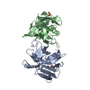
 Metallosphaera sedula (archaea)
 X-RAY DIFFRACTION /
  Authors
 Citation
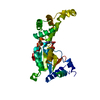
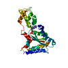
 Structure visualization
 Downloads & links
 Other downloads


 PDBj
PDBj


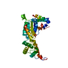
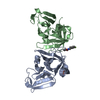
 Assembly
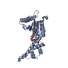
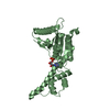


 Mass: 427.201 Da / Num. of mol.: 3 / Source method: obtained synthetically / Formula: C10H15N5O10P2 / Comment: ADP, energy-carrying molecule*YM
 Mass: 18.015 Da / Num. of mol.: 12 / Source method: isolated from a natural source / Formula: H2O
 Sample preparation
 / Beamline: ID23-2 / Wavelength: 0.8726
  Processing