 Movie
Movie Controller

[English] 日本語
 Yorodumi
- PDB-5drz: Crystal structure of anti-HIV-1 antibody F240 Fab in complex with... -
+ Open data

- Basic information
| Entry | Database: PDB / ID: 5drz | ||||||
|---|---|---|---|---|---|---|---|
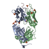
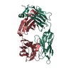
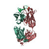
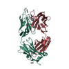
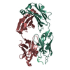
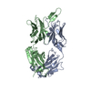
| Title | Crystal structure of anti-HIV-1 antibody F240 Fab in complex with gp41 peptide | ||||||
 Components Components |
| ||||||
 Keywords Keywords | IMMUNE SYSTEM / IgG / VIRAL PROTEIN / HIV gp41 ENVELOPE PROTEIN | ||||||
| Function / homology |  Function and homology information Function and homology information: / Dectin-2 family / positive regulation of plasma membrane raft polarization / positive regulation of receptor clustering / host cell endosome membrane / clathrin-dependent endocytosis of virus by host cell / viral protein processing / fusion of virus membrane with host plasma membrane / fusion of virus membrane with host endosome membrane / viral envelope ...: / Dectin-2 family / positive regulation of plasma membrane raft polarization / positive regulation of receptor clustering / host cell endosome membrane / clathrin-dependent endocytosis of virus by host cell / viral protein processing / fusion of virus membrane with host plasma membrane / fusion of virus membrane with host endosome membrane / viral envelope / virion attachment to host cell / host cell plasma membrane / virion membrane / structural molecule activity / membrane Similarity search - Function | ||||||
| Biological species |  Homo sapiens (human) Homo sapiens (human) HIV-1 M:B_ARV2/SF2 (virus) HIV-1 M:B_ARV2/SF2 (virus) | ||||||
| Method |  X-RAY DIFFRACTION / SYNCHROTRON / MOLECULAR REPLACEMENT / Resolution: 2.54 Å X-RAY DIFFRACTION / SYNCHROTRON / MOLECULAR REPLACEMENT / Resolution: 2.54 Å | ||||||
 Authors Authors | Gohain, N. / Tolbert, W.D. / Pazgier, M. | ||||||
| Funding support |  United States, 1items United States, 1items
| ||||||
 Citation Citation | Journal: Sci Rep / Year: 2016 Title: Molecular basis for epitope recognition by non-neutralizing anti-gp41 antibody F240. Authors: Gohain, N. / Tolbert, W.D. / Orlandi, C. / Richard, J. / Ding, S. / Chen, X. / Bonsor, D.A. / Sundberg, E.J. / Lu, W. / Ray, K. / Finzi, A. / Lewis, G.K. / Pazgier, M. | ||||||
| History |
|
- Structure visualization
| Structure viewer | Molecule: MolmilJmol/JSmol |
|---|
- Downloads & links
-Download
| PDBx/mmCIF format | 5drz.cif.gz | 361.8 KB | Display | PDBx/mmCIF format |
|---|---|---|---|---|
| PDB format | pdb5drz.ent.gz | 296.1 KB | Display | PDB format |
| PDBx/mmJSON format | 5drz.json.gz | Tree view | PDBx/mmJSON format | |
| Others |  Other downloads Other downloads |
-Validation report
| Arichive directory | https://data.pdbj.org/pub/pdb/validation_reports/dr/5drzftp://data.pdbj.org/pub/pdb/validation_reports/dr/5drz | HTTPS FTP |
|---|
-Related structure data
| Related structure data |  3tnnS S: Starting model for refinement |
|---|---|
| Similar structure data |


-Links
 PDBj
PDBj


- Assembly
| Deposited unit | 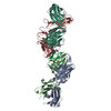
| ||||||||||||
|---|---|---|---|---|---|---|---|---|---|---|---|---|---|
| 1 | 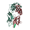
| ||||||||||||
| 2 | 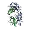
| ||||||||||||
| Unit cell |
| ||||||||||||
| Noncrystallographic symmetry (NCS) | NCS oper:
|
-Components
| #1: Antibody | Mass: 24363.109 Da / Num. of mol.: 2 Source method: isolated from a genetically manipulated source Source: (gene. exp.) Homo sapiens (human) / Cell line (production host): HEK-293 / Production host: Homo sapiens (human)#2: Antibody | Mass: 24970.748 Da / Num. of mol.: 2 Source method: isolated from a genetically manipulated source Source: (gene. exp.) Homo sapiens (human) / Cell line (production host): HEK-293 / Production host: Homo sapiens (human)#3: Protein/peptide | Mass: 4085.668 Da / Num. of mol.: 2 / Source method: obtained synthetically / Source: (synth.) HIV-1 M:B_ARV2/SF2 (virus) / References: UniProt: P03378#4: Chemical | ChemComp-MG / |   Mass: 24.305 Da / Num. of mol.: 1 / Source method: obtained synthetically / Formula: Mg Mass: 24.305 Da / Num. of mol.: 1 / Source method: obtained synthetically / Formula: Mg#5: Water | ChemComp-HOH / |  Mass: 18.015 Da / Num. of mol.: 153 / Source method: isolated from a natural source / Formula: H2O Mass: 18.015 Da / Num. of mol.: 153 / Source method: isolated from a natural source / Formula: H2OHas protein modification | Y | |
|---|
-Experimental details
-Experiment
| Experiment | Method: X-RAY DIFFRACTION |
|---|
- Sample preparation
| Crystal | Density Matthews: 2.52 Å3/Da / Density % sol: 51.2 % |
|---|---|
| Crystal grow | Temperature: 294 K / Method: vapor diffusion, hanging drop / pH: 4.5 Details: 25% PEG 3350, 15% Isopropanol, 0.2M Ammonium citrate pH4.5 PH range: 4.0-5.0 |
-Data collection
| Diffraction | Mean temperature: 100 K |
|---|---|
| Diffraction source | Source: SYNCHROTRON / Site: SSRL / Beamline: BL7-1 / Wavelength: 0.9753 Å |
| Detector | Type: ADSC QUANTUM 315 / Detector: CCD / Date: May 4, 2013 / Details: RH coated flat mirror |
| Radiation | Monochromator: Si(111) / Protocol: SINGLE WAVELENGTH / Monochromatic (M) / Laue (L): M / Scattering type: x-ray |
| Radiation wavelength | Wavelength: 0.9753 Å / Relative weight: 1 |
| Reflection | Resolution: 2.54→168.79 Å / Num. all: 33289 / Num. obs: 33289 / % possible obs: 98.8 % / Observed criterion σ(F): 0 / Redundancy: 2.9 % / Rmerge(I) obs: 0.158 / Net I/σ(I): 9.4 |
| Reflection shell | Resolution: 2.54→2.58 Å / Redundancy: 2.9 % / Rmerge(I) obs: 0.957 / Mean I/σ(I) obs: 1.4 / % possible all: 98.6 |
- Processing
| Software |
| ||||||||||||||||||||||||||||||||||||||||||||||||||||||||||||||||||||||||||||||||||||||||||||||||||||||||||||||||||||||||||||||||||||||||||||||||||||||||||||||||||||||||||||||||||||||
|---|---|---|---|---|---|---|---|---|---|---|---|---|---|---|---|---|---|---|---|---|---|---|---|---|---|---|---|---|---|---|---|---|---|---|---|---|---|---|---|---|---|---|---|---|---|---|---|---|---|---|---|---|---|---|---|---|---|---|---|---|---|---|---|---|---|---|---|---|---|---|---|---|---|---|---|---|---|---|---|---|---|---|---|---|---|---|---|---|---|---|---|---|---|---|---|---|---|---|---|---|---|---|---|---|---|---|---|---|---|---|---|---|---|---|---|---|---|---|---|---|---|---|---|---|---|---|---|---|---|---|---|---|---|---|---|---|---|---|---|---|---|---|---|---|---|---|---|---|---|---|---|---|---|---|---|---|---|---|---|---|---|---|---|---|---|---|---|---|---|---|---|---|---|---|---|---|---|---|---|---|---|---|---|
| Refinement | Method to determine structure: MOLECULAR REPLACEMENT Starting model: 3TNN Resolution: 2.54→168.79 Å / Cor.coef. Fo:Fc: 0.94 / Cor.coef. Fo:Fc free: 0.894 / SU B: 29.977 / SU ML: 0.298 / Cross valid method: THROUGHOUT / ESU R: 0.996 / ESU R Free: 0.325 / Stereochemistry target values: MAXIMUM LIKELIHOOD / Details: HYDROGENS HAVE BEEN ADDED IN THE RIDING POSITIONS
| ||||||||||||||||||||||||||||||||||||||||||||||||||||||||||||||||||||||||||||||||||||||||||||||||||||||||||||||||||||||||||||||||||||||||||||||||||||||||||||||||||||||||||||||||||||||
| Solvent computation | Ion probe radii: 0.8 Å / Shrinkage radii: 0.8 Å / VDW probe radii: 1.2 Å / Solvent model: MASK | ||||||||||||||||||||||||||||||||||||||||||||||||||||||||||||||||||||||||||||||||||||||||||||||||||||||||||||||||||||||||||||||||||||||||||||||||||||||||||||||||||||||||||||||||||||||
| Displacement parameters | Biso mean: 43.223 Å2
| ||||||||||||||||||||||||||||||||||||||||||||||||||||||||||||||||||||||||||||||||||||||||||||||||||||||||||||||||||||||||||||||||||||||||||||||||||||||||||||||||||||||||||||||||||||||
| Refinement step | Cycle: LAST / Resolution: 2.54→168.79 Å
| ||||||||||||||||||||||||||||||||||||||||||||||||||||||||||||||||||||||||||||||||||||||||||||||||||||||||||||||||||||||||||||||||||||||||||||||||||||||||||||||||||||||||||||||||||||||
| Refine LS restraints |
|