 Movie
Movie Controller

[English] 日本語
 Yorodumi
- PDB-1cu4: CRYSTAL STRUCTURE OF THE ANTI-PRION FAB 3F4 IN COMPLEX WITH ITS P... -
+ Open data

- Basic information
| Entry | Database: PDB / ID: 1cu4 | ||||||
|---|---|---|---|---|---|---|---|
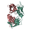
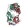
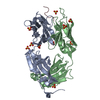
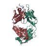
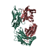
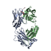
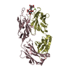
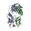
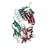
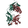
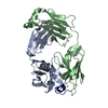
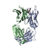
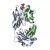
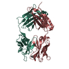
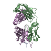
| Title | CRYSTAL STRUCTURE OF THE ANTI-PRION FAB 3F4 IN COMPLEX WITH ITS PEPTIDE EPITOPE | ||||||
 Components Components |
| ||||||
 Keywords Keywords | IMMUNE SYSTEM / IMMUNOGLOBULIN FOLD | ||||||
| Function / homology | Immunoglobulins / Immunoglobulin-like / Sandwich / Mainly Beta Function and homology information Function and homology information | ||||||
| Biological species |  | ||||||
| Method |  X-RAY DIFFRACTION / Resolution: 2.9 Å X-RAY DIFFRACTION / Resolution: 2.9 Å | ||||||
 Authors Authors | Kanyo, Z.F. / Pan, K.M. / Williamson, R.A. / Burton, D.R. / Prusiner, S.B. / Fletterick, R.J. / Cohen, F.E. | ||||||
 Citation Citation | Journal: J.Mol.Biol. / Year: 1999 Title: Antibody binding defines a structure for an epitope that participates in the PrPC-->PrPSc conformational change. Authors: Kanyo, Z.F. / Pan, K.M. / Williamson, R.A. / Burton, D.R. / Prusiner, S.B. / Fletterick, R.J. / Cohen, F.E. | ||||||
| History |
|
- Structure visualization
| Structure viewer | Molecule: MolmilJmol/JSmol |
|---|
- Downloads & links
-Download
| PDBx/mmCIF format | 1cu4.cif.gz | 89.5 KB | Display | PDBx/mmCIF format |
|---|---|---|---|---|
| PDB format | pdb1cu4.ent.gz | 68.9 KB | Display | PDB format |
| PDBx/mmJSON format | 1cu4.json.gz | Tree view | PDBx/mmJSON format | |
| Others |  Other downloads Other downloads |
-Validation report
| Arichive directory | https://data.pdbj.org/pub/pdb/validation_reports/cu/1cu4ftp://data.pdbj.org/pub/pdb/validation_reports/cu/1cu4 | HTTPS FTP |
|---|
-Related structure data


-Links
 PDBj
PDBj

- Assembly
| Deposited unit | 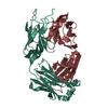
| ||||||||||
|---|---|---|---|---|---|---|---|---|---|---|---|
| 1 |
| ||||||||||
| Unit cell |
|
-Components
| #1: Antibody | Mass: 24080.775 Da / Num. of mol.: 1 / Fragment: FAB ANTIBODY 3F4, LIGHT CHAIN / Source method: isolated from a natural source / Source: (natural) |
|---|---|
| #2: Antibody | Mass: 23342.943 Da / Num. of mol.: 1 / Fragment: FAB ANTIBODY 3F4, HEAVY CHAIN / Source method: isolated from a natural source / Source: (natural) |
| #3: Protein/peptide | Mass: 1131.390 Da / Num. of mol.: 1 / Source method: obtained synthetically Details: sequence cooresponding to aa104-113 of Syrian hamster prion protein |
| #4: Water | ChemComp-HOH /  Mass: 18.015 Da / Num. of mol.: 9 / Source method: isolated from a natural source / Formula: H2O Mass: 18.015 Da / Num. of mol.: 9 / Source method: isolated from a natural source / Formula: H2O |
| Has protein modification | Y |
-Experimental details
-Experiment
| Experiment | Method: X-RAY DIFFRACTION / Number of used crystals: 1 |
|---|
- Sample preparation
| Crystal | Density Matthews: 2.11 Å3/Da / Density % sol: 41.61 % | ||||||||||||||||||||||||||||||
|---|---|---|---|---|---|---|---|---|---|---|---|---|---|---|---|---|---|---|---|---|---|---|---|---|---|---|---|---|---|---|---|
| Crystal grow | Temperature: 298 K / Method: vapor diffusion, hanging drop / pH: 7 Details: PEG 8000, pH 7.0, VAPOR DIFFUSION, HANGING DROP at 298K, temperature 298.0K | ||||||||||||||||||||||||||||||
| Crystal grow | *PLUS | ||||||||||||||||||||||||||||||
| Components of the solutions | *PLUS
|
-Data collection
| Diffraction | Mean temperature: 298 K |
|---|---|
| Diffraction source | Source: ROTATING ANODE / Type: RIGAKU RU200 / Wavelength: 1.5418 |
| Detector | Type: RIGAKU RAXIS IIC / Detector: IMAGE PLATE / Date: May 28, 1997 |
| Radiation | Protocol: SINGLE WAVELENGTH / Monochromatic (M) / Laue (L): M / Scattering type: x-ray |
| Radiation wavelength | Wavelength: 1.5418 Å / Relative weight: 1 |
| Reflection | Resolution: 2.9→20 Å / Num. all: 8689 / Num. obs: 8689 / % possible obs: 92.1 % / Observed criterion σ(I): 2 / Redundancy: 3.3 % / Biso Wilson estimate: 36.2 Å2 / Rmerge(I) obs: 0.077 / Net I/σ(I): 9.5 |
| Reflection shell | Resolution: 2.9→3.03 Å / Redundancy: 2.9 % / Rmerge(I) obs: 0.216 / % possible all: 90.4 |
| Reflection shell | *PLUS % possible obs: 90.4 % / Num. unique obs: 1011 |
- Processing
| Software |
| ||||||||||||||||||||
|---|---|---|---|---|---|---|---|---|---|---|---|---|---|---|---|---|---|---|---|---|---|
| Refinement | Resolution: 2.9→20 Å / Cross valid method: THROUGHOUT / σ(F): 2 / Stereochemistry target values: Engh & Huber
| ||||||||||||||||||||
| Refinement step | Cycle: LAST / Resolution: 2.9→20 Å
| ||||||||||||||||||||
| Refine LS restraints |
|
