 Movie
Movie Controller

[English] 日本語
 Yorodumi
- PDB-4odv: Fab Structure of lipid A-specific antibody A6 in complex with lip... -
+ Open data

- Basic information
| Entry | Database: PDB / ID: 4odv | |||||||||
|---|---|---|---|---|---|---|---|---|---|---|
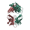
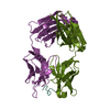
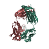
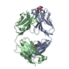
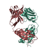
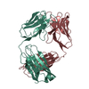
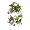
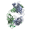
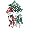
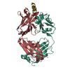
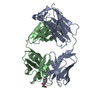
| Title | Fab Structure of lipid A-specific antibody A6 in complex with lipid A carbohydrate backbone | |||||||||
 Components Components |
| |||||||||
 Keywords Keywords | IMMUNE SYSTEM / Carbohydrate binding | |||||||||
| Function / homology | Immunoglobulins / Immunoglobulin-like / Sandwich / Mainly Beta / DI(HYDROXYETHYL)ETHER Function and homology information Function and homology information | |||||||||
| Biological species |  | |||||||||
| Method |  X-RAY DIFFRACTION / SYNCHROTRON / MOLECULAR REPLACEMENT / molecular replacement / Resolution: 2.15 Å X-RAY DIFFRACTION / SYNCHROTRON / MOLECULAR REPLACEMENT / molecular replacement / Resolution: 2.15 Å | |||||||||
 Authors Authors | Haji-Ghassemi, O. / Evans, S.V. | |||||||||
 Citation Citation | Journal: J.Biol.Chem. / Year: 2015 Title: Structural Basis for Antibody Recognition of Lipid A: INSIGHTS TO POLYSPECIFICITY TOWARD SINGLE-STRANDED DNA. Authors: Haji-Ghassemi, O. / Muller-Loennies, S. / Rodriguez, T. / Brade, L. / Kosma, P. / Brade, H. / Evans, S.V. | |||||||||
| History |
|
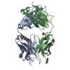
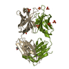
- Structure visualization
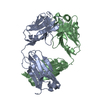
| Structure viewer | Molecule: MolmilJmol/JSmol |
|---|
- Downloads & links
-Download
| PDBx/mmCIF format | 4odv.cif.gz | 101.7 KB | Display | PDBx/mmCIF format |
|---|---|---|---|---|
| PDB format | pdb4odv.ent.gz | 76.1 KB | Display | PDB format |
| PDBx/mmJSON format | 4odv.json.gz | Tree view | PDBx/mmJSON format | |
| Others |  Other downloads Other downloads |
-Validation report
| Arichive directory | https://data.pdbj.org/pub/pdb/validation_reports/od/4odvftp://data.pdbj.org/pub/pdb/validation_reports/od/4odv | HTTPS FTP |
|---|
-Related structure data
| Related structure data | 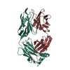 4odsC 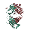 4odtSC 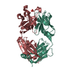 4oduC  4odwC 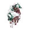 4z8fC  4z95C  4ody 4odz 4oe0 C: citing same article ( S: Starting model for refinement |
|---|---|
| Similar structure data |


-Links
 PDBj
PDBj

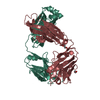
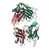
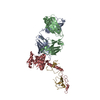
- Assembly
| Deposited unit | 
| ||||||||
|---|---|---|---|---|---|---|---|---|---|
| 1 |
| ||||||||
| Unit cell |
|
-Components
| #1: Antibody | Mass: 23851.348 Da / Num. of mol.: 1 / Source method: isolated from a natural source / Source: (natural) |
|---|---|
| #2: Antibody | Mass: 23792.738 Da / Num. of mol.: 1 / Source method: isolated from a natural source / Source: (natural) |
| #3: Polysaccharide | 2-amino-2-deoxy-4-O-phosphono-beta-D-glucopyranose-(1-6)-2-amino-2-deoxy-1-O-phosphono-alpha-D-glucopyranose Source method: isolated from a genetically manipulated source |
| #4: Chemical | ChemComp-PEG /   Mass: 106.120 Da / Num. of mol.: 1 / Source method: obtained synthetically / Formula: C4H10O3 Mass: 106.120 Da / Num. of mol.: 1 / Source method: obtained synthetically / Formula: C4H10O3 |
| #5: Water | ChemComp-HOH /  Mass: 18.015 Da / Num. of mol.: 94 / Source method: isolated from a natural source / Formula: H2O Mass: 18.015 Da / Num. of mol.: 94 / Source method: isolated from a natural source / Formula: H2O |
| Has protein modification | Y |
-Experimental details
-Experiment
| Experiment | Method: X-RAY DIFFRACTION / Number of used crystals: 1 |
|---|
- Sample preparation
| Crystal | Density Matthews: 1.95 Å3/Da / Density % sol: 37.03 % |
|---|---|
| Crystal grow | Temperature: 289 K / Method: vapor diffusion, hanging drop / pH: 7.5 Details: 0.1M HEPES pH 7.5, 25% (w/v) PEG 3350, VAPOR DIFFUSION, HANGING DROP, temperature 289K |
-Data collection
| Diffraction | Mean temperature: 100 K | |||||||||||||||||||||||||||||||||||||||||||||||||||||||||||||||||||||||||||||
|---|---|---|---|---|---|---|---|---|---|---|---|---|---|---|---|---|---|---|---|---|---|---|---|---|---|---|---|---|---|---|---|---|---|---|---|---|---|---|---|---|---|---|---|---|---|---|---|---|---|---|---|---|---|---|---|---|---|---|---|---|---|---|---|---|---|---|---|---|---|---|---|---|---|---|---|---|---|---|
| Diffraction source | Source: SYNCHROTRON / Site: CLSI  / Beamline: 08ID-1 / Wavelength: 0.9794 Å / Beamline: 08ID-1 / Wavelength: 0.9794 Å | |||||||||||||||||||||||||||||||||||||||||||||||||||||||||||||||||||||||||||||
| Detector | Type: MARMOSAIC 300 mm CCD / Detector: CCD / Date: Dec 1, 2011 / Details: Vertical focusing mirror | |||||||||||||||||||||||||||||||||||||||||||||||||||||||||||||||||||||||||||||
| Radiation | Monochromator: Double crystal monochromator / Protocol: SINGLE WAVELENGTH / Monochromatic (M) / Laue (L): M / Scattering type: x-ray | |||||||||||||||||||||||||||||||||||||||||||||||||||||||||||||||||||||||||||||
| Radiation wavelength | Wavelength: 0.9794 Å / Relative weight: 1 | |||||||||||||||||||||||||||||||||||||||||||||||||||||||||||||||||||||||||||||
| Reflection | Resolution: 2.15→25 Å / Num. obs: 21071 / % possible obs: 99.7 % / Redundancy: 4.9 % / Rmerge(I) obs: 0.085 / Χ2: 1.064 / Net I/σ(I): 12.8 | |||||||||||||||||||||||||||||||||||||||||||||||||||||||||||||||||||||||||||||
| Reflection shell |
|
-Phasing
| Phasing | Method: molecular replacement | |||||||||
|---|---|---|---|---|---|---|---|---|---|---|
| Phasing MR | Model details: Phaser MODE: MR_AUTO
|
- Processing
| Software |
| |||||||||||||||||||||||||||||||||||||||||||||||||||||||||||||||||||||||||||
|---|---|---|---|---|---|---|---|---|---|---|---|---|---|---|---|---|---|---|---|---|---|---|---|---|---|---|---|---|---|---|---|---|---|---|---|---|---|---|---|---|---|---|---|---|---|---|---|---|---|---|---|---|---|---|---|---|---|---|---|---|---|---|---|---|---|---|---|---|---|---|---|---|---|---|---|---|
| Refinement | Method to determine structure: MOLECULAR REPLACEMENT Starting model: PDB ENTRY 4ODT Resolution: 2.15→24.83 Å / Cor.coef. Fo:Fc: 0.955 / Cor.coef. Fo:Fc free: 0.93 / WRfactor Rfree: 0.2785 / WRfactor Rwork: 0.2213 / Occupancy max: 1 / Occupancy min: 0.5 / FOM work R set: 0.8231 / SU B: 6.355 / SU ML: 0.164 / SU R Cruickshank DPI: 0.3422 / SU Rfree: 0.2281 / Cross valid method: THROUGHOUT / σ(F): 0 / ESU R: 0.342 / ESU R Free: 0.228 / Stereochemistry target values: MAXIMUM LIKELIHOOD Details: HYDROGENS HAVE BEEN ADDED IN THE RIDING POSITIONS U VALUES : REFINED INDIVIDUALLY
| |||||||||||||||||||||||||||||||||||||||||||||||||||||||||||||||||||||||||||
| Solvent computation | Ion probe radii: 0.8 Å / Shrinkage radii: 0.8 Å / VDW probe radii: 1.2 Å / Solvent model: MASK | |||||||||||||||||||||||||||||||||||||||||||||||||||||||||||||||||||||||||||
| Displacement parameters | Biso max: 112.04 Å2 / Biso mean: 48.6741 Å2 / Biso min: 24.71 Å2
| |||||||||||||||||||||||||||||||||||||||||||||||||||||||||||||||||||||||||||
| Refinement step | Cycle: LAST / Resolution: 2.15→24.83 Å
| |||||||||||||||||||||||||||||||||||||||||||||||||||||||||||||||||||||||||||
| Refine LS restraints |
| |||||||||||||||||||||||||||||||||||||||||||||||||||||||||||||||||||||||||||
| LS refinement shell | Resolution: 2.151→2.207 Å / Total num. of bins used: 20
|
