 Movie
Movie Controller

[English] 日本語
 Yorodumi
- PDB-4nxq: Crystal Structure of T-cell Lymphoma Invasion and Metastasis-1 PD... -
+ Open data

- Basic information
| Entry | Database: PDB / ID: 4nxq | ||||||
|---|---|---|---|---|---|---|---|
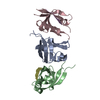
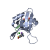
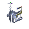
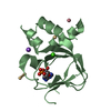
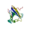
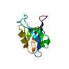
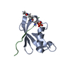
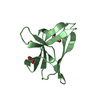
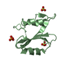
| Title | Crystal Structure of T-cell Lymphoma Invasion and Metastasis-1 PDZ Domain Quadruple Mutant (QM) in Complex With Caspr4 Peptide | ||||||
 Components Components |
| ||||||
 Keywords Keywords | signaling Protein/Peptide / Beta barrel fold protein / PDZ domain / peptide binding / specificity mutant / scaffold signaling protein for cell adhesion and cell junction / signaling domain / protein-peptide complex / signaling Protein-Peptide complex | ||||||
| Function / homology |  Function and homology information Function and homology informationregulation of dopaminergic neuron differentiation / regulation of synaptic transmission, dopaminergic / regulation of grooming behavior / Activated NTRK2 signals through CDK5 / non-canonical Wnt signaling pathway / regulation of epithelial to mesenchymal transition / cell-cell contact zone / regulation of small GTPase mediated signal transduction / Wnt signaling pathway, planar cell polarity pathway / positive regulation of axonogenesis ...regulation of dopaminergic neuron differentiation / regulation of synaptic transmission, dopaminergic / regulation of grooming behavior / Activated NTRK2 signals through CDK5 / non-canonical Wnt signaling pathway / regulation of epithelial to mesenchymal transition / cell-cell contact zone / regulation of small GTPase mediated signal transduction / Wnt signaling pathway, planar cell polarity pathway / positive regulation of axonogenesis / NRAGE signals death through JNK / small GTPase-mediated signal transduction / regulation of synaptic transmission, GABAergic / Rac protein signal transduction / CDC42 GTPase cycle / EPH-ephrin mediated repulsion of cells / RAC3 GTPase cycle / RHOA GTPase cycle / RAC2 GTPase cycle / ephrin receptor signaling pathway / positive regulation of epithelial to mesenchymal transition / cell projection / RAC1 GTPase cycle / EPHB-mediated forward signaling / guanyl-nucleotide exchange factor activity / cell-matrix adhesion / kinase binding / cell-cell junction / cell migration / G alpha (12/13) signalling events / protein-containing complex assembly / presynaptic membrane / cell adhesion / positive regulation of cell migration / positive regulation of cell population proliferation / lipid binding / synapse / plasma membrane / cytosol Similarity search - Function | ||||||
| Biological species |  Homo sapiens (human) Homo sapiens (human) | ||||||
| Method |  X-RAY DIFFRACTION / SYNCHROTRON / MOLECULAR REPLACEMENT / Resolution: 2.1 Å X-RAY DIFFRACTION / SYNCHROTRON / MOLECULAR REPLACEMENT / Resolution: 2.1 Å | ||||||
 Authors Authors | Liu, X. / Speckhard, D.C. / Shepherd, T.R. / Hengel, S.R. / Fuentes, E.J. | ||||||
 Citation Citation | Journal: Structure / Year: 2016 Title: Distinct Roles for Conformational Dynamics in Protein-Ligand Interactions. Authors: Liu, X. / Speckhard, D.C. / Shepherd, T.R. / Sun, Y.J. / Hengel, S.R. / Yu, L. / Fowler, C.A. / Gakhar, L. / Fuentes, E.J. | ||||||
| History |
|
- Structure visualization
| Structure viewer | Molecule: MolmilJmol/JSmol |
|---|
- Downloads & links
-Download
| PDBx/mmCIF format | 4nxq.cif.gz | 72 KB | Display | PDBx/mmCIF format |
|---|---|---|---|---|
| PDB format | pdb4nxq.ent.gz | 54.4 KB | Display | PDB format |
| PDBx/mmJSON format | 4nxq.json.gz | Tree view | PDBx/mmJSON format | |
| Others |  Other downloads Other downloads |
-Validation report
| Arichive directory | https://data.pdbj.org/pub/pdb/validation_reports/nx/4nxqftp://data.pdbj.org/pub/pdb/validation_reports/nx/4nxq | HTTPS FTP |
|---|
-Related structure data
| Related structure data | 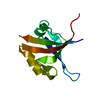 4nxpC 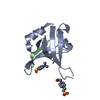 4nxrC 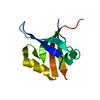 3kzdS S: Starting model for refinement C: citing same article ( |
|---|---|
| Similar structure data |


-Links
 PDBj
PDBj


- Assembly
| Deposited unit | 
| ||||||||
|---|---|---|---|---|---|---|---|---|---|
| 1 | 
| ||||||||
| 2 | 
| ||||||||
| 3 | 
| ||||||||
| Unit cell |
|
-Components
| #1: Protein | Mass: 10192.402 Da / Num. of mol.: 3 / Fragment: PDZ domain / Mutation: L911M, K912E, L915F, L920V Source method: isolated from a genetically manipulated source Source: (gene. exp.) Homo sapiens (human) / Strain: human / Gene: TIAM1 / Plasmid: PET21A-6HIS-rTEV / Production host:  #2: Protein/peptide | Mass: 1105.176 Da / Num. of mol.: 3 / Fragment: UNP residues 1301-1308 / Source method: obtained synthetically / Source: (synth.) Homo sapiens (human) / References: UniProt: Q9C0A0#3: Water | ChemComp-HOH / |  Mass: 18.015 Da / Num. of mol.: 160 / Source method: isolated from a natural source / Formula: H2O Mass: 18.015 Da / Num. of mol.: 160 / Source method: isolated from a natural source / Formula: H2O |
|---|
-Experimental details
-Experiment
| Experiment | Method: X-RAY DIFFRACTION / Number of used crystals: 1 |
|---|
- Sample preparation
| Crystal | Density Matthews: 2.03 Å3/Da / Density % sol: 39.39 % |
|---|---|
| Crystal grow | Temperature: 291 K / Method: vapor diffusion, hanging drop / pH: 6.8 Details: 0.1M Magnesium chloride, 0.1M MES, pH=6.5, 20% PEG 4000, pH 6.8, VAPOR DIFFUSION, HANGING DROP, temperature 291K |
-Data collection
| Diffraction | Mean temperature: 100 K |
|---|---|
| Diffraction source | Source: SYNCHROTRON / Site: ALS  / Beamline: 4.2.2 / Wavelength: 1 Å / Beamline: 4.2.2 / Wavelength: 1 Å |
| Detector | Type: NOIR-1 / Detector: CCD / Date: Dec 18, 2010 |
| Radiation | Monochromator: double crystal Si(111) / Protocol: SINGLE WAVELENGTH / Monochromatic (M) / Laue (L): M / Scattering type: x-ray |
| Radiation wavelength | Wavelength: 1 Å / Relative weight: 1 |
| Reflection | Resolution: 2.1→37.52 Å / Num. all: 16019 / Num. obs: 15923 / % possible obs: 99.4 % / Observed criterion σ(F): 0 / Observed criterion σ(I): 0 / Redundancy: 3.7 % / Biso Wilson estimate: 22.85 Å2 / Rmerge(I) obs: 0.081 / Net I/σ(I): 13.9 |
| Reflection shell | Resolution: 2.1→2.21 Å / Redundancy: 3.8 % / Rmerge(I) obs: 0.383 / Mean I/σ(I) obs: 3.9 / Num. unique all: 2334 / % possible all: 100 |
- Processing
| Software |
| |||||||||||||||||||||||||||||||||||||||||||||||||
|---|---|---|---|---|---|---|---|---|---|---|---|---|---|---|---|---|---|---|---|---|---|---|---|---|---|---|---|---|---|---|---|---|---|---|---|---|---|---|---|---|---|---|---|---|---|---|---|---|---|---|
| Refinement | Method to determine structure: MOLECULAR REPLACEMENT Starting model: PDB entry 3KZD Resolution: 2.1→37.52 Å / SU ML: 0.23 / Cross valid method: THROUGHOUT / σ(F): 1.34 / Phase error: 23.61 / Stereochemistry target values: MAXIMUM LIKELIHOOD
| |||||||||||||||||||||||||||||||||||||||||||||||||
| Solvent computation | Shrinkage radii: 0.9 Å / VDW probe radii: 1.11 Å / Solvent model: FLAT BULK SOLVENT MODEL | |||||||||||||||||||||||||||||||||||||||||||||||||
| Refinement step | Cycle: LAST / Resolution: 2.1→37.52 Å
| |||||||||||||||||||||||||||||||||||||||||||||||||
| Refine LS restraints |
| |||||||||||||||||||||||||||||||||||||||||||||||||
| LS refinement shell |
|
