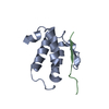
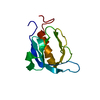
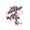
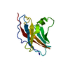
[English] 日本語
Yorodumi
- PDB-4gvc: Crystal Structure of T-cell Lymphoma Invasion and Metastasis-1 PD... -

+
Open data


ID or keywords:

Loading...

-
Basic information

Entry
Database: PDB / ID: 4gvc
TitleCrystal Structure of T-cell Lymphoma Invasion and Metastasis-1 PDZ in complex with phosphorylated Syndecan1 Peptide
Components
  • Syndecan-1Syndecan 1
  • T-lymphoma invasion and metastasis-inducing protein 1
KeywordsSIGNALING PROTEIN / phosphorylation / peptide conformational change / new binding pocket / scaffold signaling protein for cell adhesion and cell junction / syndecan1 P1 Tyr phosphorylation / sydencan1 N-terminal Thr dansylation
Function / homology
Function and homology information


myoblast development / positive regulation of Schwann cell chemotaxis / striated muscle cell development / regulation of non-canonical Wnt signaling pathway / regulation of dopaminergic neuron differentiation / Defective B3GALT6 causes EDSP2 and SEMDJL1 / Defective B4GALT7 causes EDS, progeroid type / Defective B3GAT3 causes JDSSDHD / Defective EXT2 causes exostoses 2 / Defective EXT1 causes exostoses 1, TRPS2 and CHDS ...myoblast development / positive regulation of Schwann cell chemotaxis / striated muscle cell development / regulation of non-canonical Wnt signaling pathway / regulation of dopaminergic neuron differentiation / Defective B3GALT6 causes EDSP2 and SEMDJL1 / Defective B4GALT7 causes EDS, progeroid type / Defective B3GAT3 causes JDSSDHD / Defective EXT2 causes exostoses 2 / Defective EXT1 causes exostoses 1, TRPS2 and CHDS / A tetrasaccharide linker sequence is required for GAG synthesis / extrinsic component of postsynaptic density membrane / HS-GAG biosynthesis / HS-GAG degradation / positive regulation of extracellular exosome assembly / Activated NTRK2 signals through CDK5 / Sertoli cell development / regulation of epithelial to mesenchymal transition / positive regulation of dendritic spine morphogenesis / regulation of insulin secretion involved in cellular response to glucose stimulus / regulation of modification of postsynaptic actin cytoskeleton / cell-cell contact zone / positive regulation of exosomal secretion / protein localization to membrane / cardiac muscle hypertrophy / activation of GTPase activity / cargo receptor activity / Wnt signaling pathway, planar cell polarity pathway / regulation of small GTPase mediated signal transduction / ureteric bud development / main axon / neuron projection extension / Syndecan interactions / small GTPase-mediated signal transduction / positive regulation of axonogenesis / Other interleukin signaling / NRAGE signals death through JNK / Rac protein signal transduction / CDC42 GTPase cycle / EPH-ephrin mediated repulsion of cells / RHOA GTPase cycle / canonical Wnt signaling pathway / positive regulation of epithelial to mesenchymal transition / RAC3 GTPase cycle / RAC2 GTPase cycle / ephrin receptor signaling pathway / Retinoid metabolism and transport / axonal growth cone / response to glucocorticoid / response to cAMP / RAC1 GTPase cycle / EPHB-mediated forward signaling / receptor-mediated endocytosis / lysosomal lumen / guanyl-nucleotide exchange factor activity / cell-matrix adhesion / regulation of ERK1 and ERK2 cascade / response to cocaine / Cell surface interactions at the vascular wall / response to hydrogen peroxide / receptor tyrosine kinase binding / response to toxic substance / ruffle membrane / kinase binding / Golgi lumen / response to calcium ion / G alpha (12/13) signalling events / cell-cell junction / cell migration / positive regulation of protein binding / microtubule binding / protein-containing complex assembly / microtubule / dendritic spine / Attachment and Entry / positive regulation of cell migration / external side of plasma membrane / neuronal cell body / glutamatergic synapse / lipid binding / synapse / positive regulation of cell population proliferation / cell surface / protein-containing complex / extracellular exosome / identical protein binding / plasma membrane / cytosol
Similarity search - Function
Syndecan / Syndecan, conserved site / Syndecans signature. / TIAM1, CC-Ex domain / Tiam1/Tiam2/Protein still life / T-lymphoma invasion and metastasis CC-Ex domain / Syndecan/Neurexin domain / Syndecan domain / Neurexin/syndecan/glycophorin C / putative band 4.1 homologues' binding motif ...Syndecan / Syndecan, conserved site / Syndecans signature. / TIAM1, CC-Ex domain / Tiam1/Tiam2/Protein still life / T-lymphoma invasion and metastasis CC-Ex domain / Syndecan/Neurexin domain / Syndecan domain / Neurexin/syndecan/glycophorin C / putative band 4.1 homologues' binding motif / Guanine-nucleotide dissociation stimulator, CDC24, conserved site / Dbl homology (DH) domain signature. / Raf-like Ras-binding domain / Raf-like Ras-binding / Ras-binding domain (RBD) profile. / Raf-like Ras-binding domain / Dbl homology (DH) domain superfamily / RhoGEF domain / Guanine nucleotide exchange factor for Rho/Rac/Cdc42-like GTPases / Dbl homology (DH) domain / Dbl homology (DH) domain profile. / PDZ domain / Pdz3 Domain / PH domain / PH domain profile. / Pleckstrin homology domain. / Pleckstrin homology domain / PDZ domain / PDZ domain profile. / Domain present in PSD-95, Dlg, and ZO-1/2. / PDZ domain / PDZ superfamily / PH-like domain superfamily / Roll / Mainly Beta
Similarity search - Domain/homology
Chem-ANS / Syndecan-1 / Rho guanine nucleotide exchange factor TIAM1
Similarity search - Component
Biological speciesHomo sapiens (human)
MethodX-RAY DIFFRACTION / SYNCHROTRON / MOLECULAR REPLACEMENT / Resolution: 1.54 Å
AuthorsLiu, X. / Shepherd, T.R. / Murray, A.M. / Xu, Z. / Fuentes, E.J.
CitationJournal: Structure / Year: 2013
Title: The structure of the Tiam1 PDZ domain/ phospho-syndecan1 complex reveals a ligand conformation that modulates protein dynamics.
Authors: Liu, X. / Shepherd, T.R. / Murray, A.M. / Xu, Z. / Fuentes, E.J.
History
DepositionAug 30, 2012Deposition site: RCSB / Processing site: RCSB
Revision 1.0Mar 13, 2013Provider: repository / Type: Initial release
Revision 1.1May 27, 2015Group: Database references
Revision 1.2Sep 13, 2023Group: Data collection / Database references ...Data collection / Database references / Derived calculations / Refinement description
Category: chem_comp_atom / chem_comp_bond ...chem_comp_atom / chem_comp_bond / database_2 / pdbx_initial_refinement_model / struct_conn / struct_ref_seq_dif / struct_site
Item: _database_2.pdbx_DOI / _database_2.pdbx_database_accession ..._database_2.pdbx_DOI / _database_2.pdbx_database_accession / _struct_conn.pdbx_dist_value / _struct_conn.pdbx_leaving_atom_flag / _struct_conn.ptnr1_auth_comp_id / _struct_conn.ptnr1_auth_seq_id / _struct_conn.ptnr1_label_atom_id / _struct_conn.ptnr1_label_comp_id / _struct_conn.ptnr1_label_seq_id / _struct_conn.ptnr2_auth_comp_id / _struct_conn.ptnr2_auth_seq_id / _struct_conn.ptnr2_label_asym_id / _struct_conn.ptnr2_label_atom_id / _struct_conn.ptnr2_label_comp_id / _struct_conn.ptnr2_label_seq_id / _struct_ref_seq_dif.details / _struct_site.pdbx_auth_asym_id / _struct_site.pdbx_auth_comp_id / _struct_site.pdbx_auth_seq_id
Revision 1.3Dec 6, 2023Group: Data collection / Category: chem_comp_atom / chem_comp_bond / Item: _chem_comp_atom.atom_id / _chem_comp_bond.atom_id_2

-
Structure visualization

Structure viewerMolecule:
MolmilJmol/JSmol

Downloads & links

-
Assembly

Deposited unit
A: T-lymphoma invasion and metastasis-inducing protein 1
B: Syndecan-1
hetero molecules


Theoretical massNumber of molelcules
Total (without water)11,5966
Polymers11,2512
Non-polymers3454
Water1,72996
1


  • Idetical with deposited unit
  • defined by author&software
TypeNameSymmetry operationNumber
identity operation1_555x,y,z1
Buried area1840 Å2
ΔGint-32 kcal/mol
Surface area6530 Å2
MethodPISA
Unit cell
Length a, b, c (Å)26.717, 50.135, 57.712
Angle α, β, γ (deg.)90.00, 90.00, 90.00
Int Tables number19
Space group name H-MP212121

-
Components

-
Protein / Protein/peptide , 2 types, 2 molecules AB

#1: Protein T-lymphoma invasion and metastasis-inducing protein 1 / TIAM-1


Mass: 10154.440 Da / Num. of mol.: 1 / Fragment: PDZ domain (UNP residues 841-930)
Source method: isolated from a genetically manipulated source
Source: (gene. exp.) Homo sapiens (human) / Strain: human / Gene: TIAM1 / Plasmid: PET21A-6HIS-rTEV / Production host: Escherichia coli (E. coli) / Strain (production host): BL21(DE3) / References: UniProt: Q13009
#2: Protein/peptide Syndecan-1 / Syndecan 1 / SYND1


Mass: 1096.060 Da / Num. of mol.: 1 / Fragment: UNP residues 303-310 / Source method: obtained synthetically / Source: (synth.) Homo sapiens (human) / References: UniProt: P18827

-
Non-polymers , 4 types, 100 molecules

#3: Chemical ChemComp-CL / CHLORIDE ION / Chloride


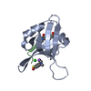
Mass: 35.453 Da / Num. of mol.: 2 / Source method: obtained synthetically / Formula: Cl
#4: Chemical ChemComp-ANS / 5-(DIMETHYLAMINO)-1-NAPHTHALENESULFONIC ACID(DANSYL ACID) / DANSYL ACID


Mass: 251.302 Da / Num. of mol.: 1 / Source method: obtained synthetically / Formula: C12H13NO3S
#5: Chemical ChemComp-NA / SODIUM ION


Mass: 22.990 Da / Num. of mol.: 1 / Source method: obtained synthetically / Formula: Na
#6: Water ChemComp-HOH / water / Water


Mass: 18.015 Da / Num. of mol.: 96 / Source method: isolated from a natural source / Formula: H2O

-
Details

Sequence detailsQ844H IS A NATURAL VARIANT.

-
Experimental details

-
Experiment

ExperimentMethod: X-RAY DIFFRACTION / Number of used crystals: 1

-
Sample preparation

CrystalDensity Matthews: 1.72 Å3/Da / Density % sol: 28.39 %
Crystal growTemperature: 291 K / Method: vapor diffusion, hanging drop / pH: 6.8
Details: 0.1 M sodium acetate, 25% PEG4000, 8% isopropanol, pH 6.8, VAPOR DIFFUSION, HANGING DROP, temperature 291K

-
Data collection

DiffractionMean temperature: 100 K
Diffraction sourceSource: SYNCHROTRON / Site: ALS / Beamline: 4.2.2 / Wavelength: 1 Å
DetectorType: NOIR-1 / Detector: CCD / Date: Apr 30, 2010
RadiationMonochromator: double crystal Si(111) / Protocol: SINGLE WAVELENGTH / Monochromatic (M) / Laue (L): M / Scattering type: x-ray
Radiation wavelengthWavelength: 1 Å / Relative weight: 1
ReflectionResolution: 1.54→37.85 Å / Num. all: 11979 / Num. obs: 11440 / % possible obs: 95.5 % / Observed criterion σ(F): 0 / Observed criterion σ(I): -3 / Redundancy: 6.4 % / Biso Wilson estimate: 13 Å2 / Rmerge(I) obs: 0.03 / Net I/σ(I): 34.4
Reflection shellResolution: 1.54→1.62 Å / Redundancy: 3.8 % / Rmerge(I) obs: 0.121 / Mean I/σ(I) obs: 9.3 / Num. unique all: 1685 / % possible all: 74.6

-
Processing

Software
NameVersionClassification
StructureStudiodata collection
PHASERphasing
REFMAC5.5.0109refinement
XDSdata reduction
SCALAdata scaling
RefinementMethod to determine structure: MOLECULAR REPLACEMENT
Starting model: PDB ENTRY 3KZD
Resolution: 1.54→37.85 Å / Cor.coef. Fo:Fc: 0.959 / Cor.coef. Fo:Fc free: 0.944 / SU B: 2.964 / SU ML: 0.055 / Cross valid method: THROUGHOUT / σ(F): 0 / ESU R: 0.138 / ESU R Free: 0.091 / Stereochemistry target values: MAXIMUM LIKELIHOOD
Details: HYDROGENS HAVE BEEN ADDED IN THE RIDING POSITIONS U VALUES : REFINED INDIVIDUALLY
RfactorNum. reflection% reflectionSelection details
Rfree0.19964 546 4.8 %RANDOM
Rwork0.16551 ---
all0.16717 10862 --
obs0.16717 10862 95.04 %-
Solvent computationIon probe radii: 0.8 Å / Shrinkage radii: 0.8 Å / VDW probe radii: 1.4 Å / Solvent model: MASK
Displacement parametersBiso mean: 13.551 Å2
Baniso -1Baniso -2Baniso -3
1-0.11 Å20 Å20 Å2
2---0.08 Å20 Å2
3----0.03 Å2
Refinement stepCycle: LAST / Resolution: 1.54→37.85 Å
ProteinNucleic acidLigandSolventTotal
Num. atoms787 0 19 96 902
Refine LS restraints
Refine-IDTypeDev idealDev ideal targetNumber
X-RAY DIFFRACTIONr_bond_refined_d0.0080.022810
X-RAY DIFFRACTIONr_bond_other_d
X-RAY DIFFRACTIONr_angle_refined_deg1.2652.0131091
X-RAY DIFFRACTIONr_angle_other_deg
X-RAY DIFFRACTIONr_dihedral_angle_1_deg6.2315100
X-RAY DIFFRACTIONr_dihedral_angle_2_deg41.6932534
X-RAY DIFFRACTIONr_dihedral_angle_3_deg12.19515145
X-RAY DIFFRACTIONr_dihedral_angle_4_deg17.547154
X-RAY DIFFRACTIONr_chiral_restr0.0760.2122
X-RAY DIFFRACTIONr_gen_planes_refined0.0050.02593
X-RAY DIFFRACTIONr_gen_planes_other
X-RAY DIFFRACTIONr_nbd_refined
X-RAY DIFFRACTIONr_nbd_other
X-RAY DIFFRACTIONr_nbtor_refined
X-RAY DIFFRACTIONr_nbtor_other
X-RAY DIFFRACTIONr_xyhbond_nbd_refined
X-RAY DIFFRACTIONr_xyhbond_nbd_other
X-RAY DIFFRACTIONr_metal_ion_refined
X-RAY DIFFRACTIONr_metal_ion_other
X-RAY DIFFRACTIONr_symmetry_vdw_refined
X-RAY DIFFRACTIONr_symmetry_vdw_other
X-RAY DIFFRACTIONr_symmetry_hbond_refined
X-RAY DIFFRACTIONr_symmetry_hbond_other
X-RAY DIFFRACTIONr_symmetry_metal_ion_refined
X-RAY DIFFRACTIONr_symmetry_metal_ion_other
X-RAY DIFFRACTIONr_mcbond_it0.921.5506
X-RAY DIFFRACTIONr_mcbond_other
X-RAY DIFFRACTIONr_mcangle_it1.6462802
X-RAY DIFFRACTIONr_scbond_it2.6483304
X-RAY DIFFRACTIONr_scangle_it4.1744.5289
X-RAY DIFFRACTIONr_rigid_bond_restr1.1113804
X-RAY DIFFRACTIONr_sphericity_free
X-RAY DIFFRACTIONr_sphericity_bonded
LS refinement shellResolution: 1.54→1.581 Å / Total num. of bins used: 20
RfactorNum. reflection% reflection
Rfree0.246 35 -
Rwork0.163 497 -
obs-532 60.73 %

+
About Yorodumi

-
News

-
Feb 9, 2022. New format data for meta-information of EMDB entries

New format data for meta-information of EMDB entries

  • Version 3 of the EMDB header file is now the official format.
  • The previous official version 1.9 will be removed from the archive.

Related info.:EMDB header

External links:wwPDB to switch to version 3 of the EMDB data model

-
Aug 12, 2020. Covid-19 info

Covid-19 info

URL: https://pdbj.org/emnavi/covid19.php

New page: Covid-19 featured information page in EM Navigator.

Related info.:Covid-19 info / Mar 5, 2020. Novel coronavirus structure data

+
Mar 5, 2020. Novel coronavirus structure data

Novel coronavirus structure data

Related info.:Yorodumi Speices / Aug 12, 2020. Covid-19 info

External links:COVID-19 featured content - PDBj / Molecule of the Month (242):Coronavirus Proteases

+
Jan 31, 2019. EMDB accession codes are about to change! (news from PDBe EMDB page)

EMDB accession codes are about to change! (news from PDBe EMDB page)

  • The allocation of 4 digits for EMDB accession codes will soon come to an end. Whilst these codes will remain in use, new EMDB accession codes will include an additional digit and will expand incrementally as the available range of codes is exhausted. The current 4-digit format prefixed with “EMD-” (i.e. EMD-XXXX) will advance to a 5-digit format (i.e. EMD-XXXXX), and so on. It is currently estimated that the 4-digit codes will be depleted around Spring 2019, at which point the 5-digit format will come into force.
  • The EM Navigator/Yorodumi systems omit the EMD- prefix.

Related info.:Q: What is EMD? / ID/Accession-code notation in Yorodumi/EM Navigator

External links:EMDB Accession Codes are Changing Soon! / Contact to PDBj

+
Jul 12, 2017. Major update of PDB

Major update of PDB

  • wwPDB released updated PDB data conforming to the new PDBx/mmCIF dictionary.
  • This is a major update changing the version number from 4 to 5, and with Remediation, in which all the entries are updated.
  • In this update, many items about electron microscopy experimental information are reorganized (e.g. em_software).
  • Now, EM Navigator and Yorodumi are based on the updated data.

External links:wwPDB Remediation / Enriched Model Files Conforming to OneDep Data Standards Now Available in the PDB FTP Archive

-
Yorodumi

Thousand views of thousand structures

  • Yorodumi is a browser for structure data from EMDB, PDB, SASBDB, etc.
  • This page is also the successor to EM Navigator detail page, and also detail information page/front-end page for Omokage search.
  • The word "yorodu" (or yorozu) is an old Japanese word meaning "ten thousand". "mi" (miru) is to see.

Related info.:EMDB / PDB / SASBDB / Comparison of 3 databanks / Yorodumi Search / Aug 31, 2016. New EM Navigator & Yorodumi / Yorodumi Papers / Jmol/JSmol / Function and homology information / Changes in new EM Navigator and Yorodumi

Read more