- PDB-4gvd: Crystal Structure of T-cell Lymphoma Invasion and Metastasis-1 PD... -
+
Open data
ID or keywords:
Loading...
-
Basic information
Entry
Database: PDB / ID: 4gvd
Title
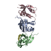
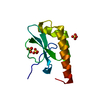
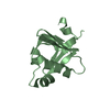
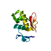
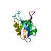
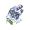
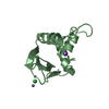
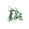
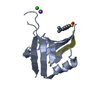
Crystal Structure of T-cell Lymphoma Invasion and Metastasis-1 PDZ in complex with Syndecan1 Peptide
Components
Syndecan-1
T-lymphoma invasion and metastasis-inducing protein 1
Keywords
SIGNALING PROTEIN / conformational change during phosphorylation / different binding pocket from phosphorylated sydencan1 / scaffold signaling protein for cell adhesion and cell junction / sydencan1 N-terminal Thr dansylation
Function / homology
Function and homology information
myoblast development / striated muscle cell development / regulation of dopaminergic neuron differentiation / Defective B3GALT6 causes EDSP2 and SEMDJL1 / Defective B4GALT7 causes EDS, progeroid type / Defective B3GAT3 causes JDSSDHD / Defective EXT2 causes exostoses 2 / Defective EXT1 causes exostoses 1, TRPS2 and CHDS / Dengue Virus Attachment and Entry / Glycosaminoglycan-protein linkage region biosynthesis ...myoblast development / striated muscle cell development / regulation of dopaminergic neuron differentiation / Defective B3GALT6 causes EDSP2 and SEMDJL1 / Defective B4GALT7 causes EDS, progeroid type / Defective B3GAT3 causes JDSSDHD / Defective EXT2 causes exostoses 2 / Defective EXT1 causes exostoses 1, TRPS2 and CHDS / Dengue Virus Attachment and Entry / Glycosaminoglycan-protein linkage region biosynthesis / HS-GAG biosynthesis / positive regulation of extracellular exosome assembly / Activated NTRK2 signals through CDK5 / non-canonical Wnt signaling pathway / Dengue Virus-Host Interactions / regulation of epithelial to mesenchymal transition / HS-GAG degradation / cell-cell contact zone / positive regulation of exosomal secretion / regulation of small GTPase mediated signal transduction / Wnt signaling pathway, planar cell polarity pathway / positive regulation of axonogenesis / Other interleukin signaling / cargo receptor activity / Syndecan interactions / NRAGE signals death through JNK / small GTPase-mediated signal transduction / RSV-host interactions / Rac protein signal transduction / CDC42 GTPase cycle / Respiratory syncytial virus (RSV) attachment and entry / EPH-ephrin mediated repulsion of cells / RAC3 GTPase cycle / RHOA GTPase cycle / RAC2 GTPase cycle / ephrin receptor signaling pathway / positive regulation of epithelial to mesenchymal transition / Retinoid metabolism and transport / RAC1 GTPase cycle / EPHB-mediated forward signaling / lysosomal lumen / receptor-mediated endocytosis / guanyl-nucleotide exchange factor activity / cell-matrix adhesion / Cell surface interactions at the vascular wall / Golgi lumen / kinase binding / cell-cell junction / cell migration / G alpha (12/13) signalling events / protein-containing complex assembly / Attachment and Entry / positive regulation of cell migration / external side of plasma membrane / positive regulation of cell population proliferation / lipid binding / synapse / cell surface / extracellular exosome / identical protein binding / plasma membrane / cytosol Similarity search - Function
A: T-lymphoma invasion and metastasis-inducing protein 1 B: T-lymphoma invasion and metastasis-inducing protein 1 C: Syndecan-1 D: Syndecan-1 hetero molecules
Resolution: 1.85→50.946 Å / Cor.coef. Fo:Fc: 0.956 / Cor.coef. Fo:Fc free: 0.943 / SU B: 3.956 / SU ML: 0.121 / Cross valid method: THROUGHOUT / ESU R: 0.184 / ESU R Free: 0.161 / Stereochemistry target values: MAXIMUM LIKELIHOOD / Details: HYDROGENS HAVE BEEN USED IF PRESENT IN THE INPUT
Rfactor
Num. reflection
% reflection
Selection details
Rfree
0.24302
655
4.9 %
RANDOM
Rwork
0.19604
-
-
-
obs
0.19848
12590
99.57 %
-
all
-
12590
-
-
Solvent computation
Ion probe radii: 0.8 Å / Shrinkage radii: 0.8 Å / VDW probe radii: 1.2 Å / Solvent model: MASK
Displacement parameters
Biso mean: 28.8 Å2
Baniso -1
Baniso -2
Baniso -3
1-
0.41 Å2
0 Å2
-0.22 Å2
2-
-
0.45 Å2
0 Å2
3-
-
-
-0.86 Å2
Refinement step
Cycle: LAST / Resolution: 1.85→50.946 Å
Protein
Nucleic acid
Ligand
Solvent
Total
Num. atoms
1428
0
37
101
1566
Refine LS restraints
Refine-ID
Type
Dev ideal
Dev ideal target
Number
X-RAY DIFFRACTION
r_bond_refined_d
0.02
0.02
1476
X-RAY DIFFRACTION
r_bond_other_d
X-RAY DIFFRACTION
r_angle_refined_deg
2.289
2.004
1986
X-RAY DIFFRACTION
r_angle_other_deg
X-RAY DIFFRACTION
r_dihedral_angle_1_deg
6.764
5
186
X-RAY DIFFRACTION
r_dihedral_angle_2_deg
43.026
23.889
54
X-RAY DIFFRACTION
r_dihedral_angle_3_deg
14.673
15
252
X-RAY DIFFRACTION
r_dihedral_angle_4_deg
18.696
15
8
X-RAY DIFFRACTION
r_chiral_restr
0.154
0.2
234
X-RAY DIFFRACTION
r_gen_planes_refined
0.01
0.02
1066
X-RAY DIFFRACTION
r_gen_planes_other
X-RAY DIFFRACTION
r_nbd_refined
X-RAY DIFFRACTION
r_nbd_other
X-RAY DIFFRACTION
r_nbtor_refined
X-RAY DIFFRACTION
r_nbtor_other
X-RAY DIFFRACTION
r_xyhbond_nbd_refined
X-RAY DIFFRACTION
r_xyhbond_nbd_other
X-RAY DIFFRACTION
r_metal_ion_refined
X-RAY DIFFRACTION
r_metal_ion_other
X-RAY DIFFRACTION
r_symmetry_vdw_refined
X-RAY DIFFRACTION
r_symmetry_vdw_other
X-RAY DIFFRACTION
r_symmetry_hbond_refined
X-RAY DIFFRACTION
r_symmetry_hbond_other
X-RAY DIFFRACTION
r_symmetry_metal_ion_refined
X-RAY DIFFRACTION
r_symmetry_metal_ion_other
X-RAY DIFFRACTION
r_mcbond_it
X-RAY DIFFRACTION
r_mcbond_other
X-RAY DIFFRACTION
r_mcangle_it
X-RAY DIFFRACTION
r_scbond_it
X-RAY DIFFRACTION
r_scangle_it
X-RAY DIFFRACTION
r_rigid_bond_restr
X-RAY DIFFRACTION
r_sphericity_free
X-RAY DIFFRACTION
r_sphericity_bonded
LS refinement shell
Resolution: 1.85→1.898 Å / Total num. of bins used: 20
Rfactor
Num. reflection
% reflection
Rfree
0.362
52
-
Rwork
0.251
905
-
obs
-
-
98.97 %
+
About Yorodumi
-
News
-
Feb 9, 2022. New format data for meta-information of EMDB entries
New format data for meta-information of EMDB entries
Version 3 of the EMDB header file is now the official format.
The previous official version 1.9 will be removed from the archive.
In the structure databanks used in Yorodumi, some data are registered as the other names, "COVID-19 virus" and "2019-nCoV". Here are the details of the virus and the list of structure data.
Jan 31, 2019. EMDB accession codes are about to change! (news from PDBe EMDB page)
EMDB accession codes are about to change! (news from PDBe EMDB page)
The allocation of 4 digits for EMDB accession codes will soon come to an end. Whilst these codes will remain in use, new EMDB accession codes will include an additional digit and will expand incrementally as the available range of codes is exhausted. The current 4-digit format prefixed with “EMD-” (i.e. EMD-XXXX) will advance to a 5-digit format (i.e. EMD-XXXXX), and so on. It is currently estimated that the 4-digit codes will be depleted around Spring 2019, at which point the 5-digit format will come into force.
The EM Navigator/Yorodumi systems omit the EMD- prefix.
Related info.:Q: What is EMD? / ID/Accession-code notation in Yorodumi/EM Navigator
Yorodumi is a browser for structure data from EMDB, PDB, SASBDB, etc.
This page is also the successor to EM Navigator detail page, and also detail information page/front-end page for Omokage search.
The word "yorodu" (or yorozu) is an old Japanese word meaning "ten thousand". "mi" (miru) is to see.
Related info.:EMDB / PDB / SASBDB / Comparison of 3 databanks / Yorodumi Search / Aug 31, 2016. New EM Navigator & Yorodumi / Yorodumi Papers / Jmol/JSmol / Function and homology information / Changes in new EM Navigator and Yorodumi
 Movie
Movie Controller

 Yorodumi
 Open data
 Basic information
 Components
 Keywords
 Function and homology information
 Homo sapiens (human)
 X-RAY DIFFRACTION /
  Authors
 Citation
 Structure visualization
 Downloads & links
 Other downloads
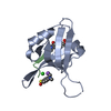


 PDBj
PDBj


 Assembly
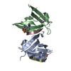


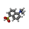

 Mass: 35.453 Da / Num. of mol.: 3 / Source method: obtained synthetically / Formula: Cl
 Mass: 22.990 Da / Num. of mol.: 2 / Source method: obtained synthetically / Formula: Na
 Mass: 251.302 Da / Num. of mol.: 2 / Source method: obtained synthetically / Formula: C12H13NO3S
 Sample preparation
 / Beamline: 4.2.2 / Wavelength: 1 Å
 Processing