 Movie
Movie Controller

[English] 日本語
 Yorodumi
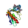
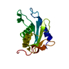
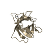
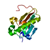
- PDB-1g2r: Structure of Cytosolic Protein of Unknown Function Coded by Gene ... -
+ Open data

- Basic information
| Entry | Database: PDB / ID: 1g2r | ||||||
|---|---|---|---|---|---|---|---|
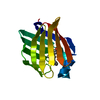
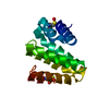
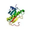
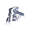
| Title | Structure of Cytosolic Protein of Unknown Function Coded by Gene from NUSA/INFB Region, a YlxR Homologue | ||||||
 Components Components | HYPOTHETICAL CYTOSOLIC PROTEIN | ||||||
 Keywords Keywords | STRUCTURAL GENOMICS / UNKNOWN FUNCTION / hypothetical / NusA-InfB operon / Streptococcus pneumoniae / PSI / Protein Structure Initiative / Midwest Center for Structural Genomics / MCSG | ||||||
| Function / homology |  Function and homology information Function and homology informationYlxR-like / YlxR domain / YlxR-like superfamily / YlxR / Protein of unknown function (DUF448) / Hypothetical Cytosolic Protein; Chain: A; / 2-Layer Sandwich / Alpha Beta Similarity search - Domain/homology | ||||||
| Biological species |   Streptococcus pneumoniae (bacteria) Streptococcus pneumoniae (bacteria) | ||||||
| Method |  X-RAY DIFFRACTION / SYNCHROTRON / MAD / Resolution: 1.35 Å X-RAY DIFFRACTION / SYNCHROTRON / MAD / Resolution: 1.35 Å | ||||||
 Authors Authors | Osipiuk, J. / Gornicki, P. / Maj, L. / Joachimiak, A. / Midwest Center for Structural Genomics (MCSG) | ||||||
 Citation Citation | Journal: Acta Crystallogr.,Sect.D / Year: 2001 Title: Streptococcus pneumonia YlxR at 1.35 A shows a putative new fold. Authors: Osipiuk, J. / Gornicki, P. / Maj, L. / Dementieva, I. / Laskowski, R. / Joachimiak, A. | ||||||
| History |
|
- Structure visualization
| Structure viewer | Molecule: MolmilJmol/JSmol |
|---|
- Downloads & links
-Download
| PDBx/mmCIF format | 1g2r.cif.gz | 59.9 KB | Display | PDBx/mmCIF format |
|---|---|---|---|---|
| PDB format | pdb1g2r.ent.gz | 43.9 KB | Display | PDB format |
| PDBx/mmJSON format | 1g2r.json.gz | Tree view | PDBx/mmJSON format | |
| Others |  Other downloads Other downloads |
-Validation report
| Arichive directory | https://data.pdbj.org/pub/pdb/validation_reports/g2/1g2rftp://data.pdbj.org/pub/pdb/validation_reports/g2/1g2r | HTTPS FTP |
|---|
-Related structure data
| Similar structure data | |
|---|---|
| Other databases |


-Links
 PDBj
PDBj- Assembly
| Deposited unit | 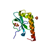
| ||||||||
|---|---|---|---|---|---|---|---|---|---|
| 1 |
| ||||||||
| Unit cell |
|
-Components
| #1: Protein | Mass: 11545.360 Da / Num. of mol.: 1 Source method: isolated from a genetically manipulated source Source: (gene. exp.) Streptococcus pneumoniae (bacteria) / Plasmid: PET15B / Species (production host): Escherichia coli / Production host: | ||
|---|---|---|---|
| #2: Chemical |   Mass: 96.063 Da / Num. of mol.: 3 / Source method: obtained synthetically / Formula: SO4 Mass: 96.063 Da / Num. of mol.: 3 / Source method: obtained synthetically / Formula: SO4#3: Water | ChemComp-HOH / |  Mass: 18.015 Da / Num. of mol.: 131 / Source method: isolated from a natural source / Formula: H2O Mass: 18.015 Da / Num. of mol.: 131 / Source method: isolated from a natural source / Formula: H2O |
-Experimental details
-Experiment
| Experiment | Method: X-RAY DIFFRACTION / Number of used crystals: 1 |
|---|
- Sample preparation
| Crystal | Density Matthews: 2.18 Å3/Da / Density % sol: 43.62 % | ||||||||||||||||||||||||||||||||||||||||||||||||
|---|---|---|---|---|---|---|---|---|---|---|---|---|---|---|---|---|---|---|---|---|---|---|---|---|---|---|---|---|---|---|---|---|---|---|---|---|---|---|---|---|---|---|---|---|---|---|---|---|---|
| Crystal grow | Temperature: 283 K / Method: vapor diffusion, hanging drop / pH: 5.6 Details: 0.2 M potassium sodium tartrate, 0.1 M tri-sodium citrate, 2.0 M ammonium sulfate, pH 5.6, VAPOR DIFFUSION, HANGING DROP, temperature 283K | ||||||||||||||||||||||||||||||||||||||||||||||||
| Crystal grow | *PLUS pH: 7.4 | ||||||||||||||||||||||||||||||||||||||||||||||||
| Components of the solutions | *PLUS
|
-Data collection
| Diffraction | Mean temperature: 100 K |
|---|---|
| Diffraction source | Source: SYNCHROTRON / Site: APS  / Beamline: 19-BM / Wavelength: 1.0332 Å / Beamline: 19-BM / Wavelength: 1.0332 Å |
| Detector | Type: CUSTOM-MADE / Detector: CCD / Date: Aug 15, 2000 |
| Radiation | Monochromator: SI 111 / Protocol: SINGLE WAVELENGTH / Monochromatic (M) / Laue (L): M / Scattering type: x-ray |
| Radiation wavelength | Wavelength: 1.0332 Å / Relative weight: 1 |
| Reflection | Resolution: 1.35→30 Å / Num. all: 21402 / Num. obs: 101345 / % possible obs: 93.5 % / Observed criterion σ(F): 1 / Observed criterion σ(I): 1 / Redundancy: 4.8 % / Biso Wilson estimate: 15.9 Å2 / Rmerge(I) obs: 0.063 / Net I/σ(I): 11.2 |
| Reflection shell | Resolution: 1.35→1.37 Å / Redundancy: 2.9 % / Rmerge(I) obs: 0.269 / Mean I/σ(I) obs: 2.7 / Num. unique all: 684 / % possible all: 61.3 |
| Reflection | *PLUS Num. obs: 21402 / Num. measured all: 101345 |
| Reflection shell | *PLUS % possible obs: 61.3 % |
- Processing
| Software |
| |||||||||||||||||||||||||
|---|---|---|---|---|---|---|---|---|---|---|---|---|---|---|---|---|---|---|---|---|---|---|---|---|---|---|
| Refinement | Method to determine structure: MAD / Resolution: 1.35→29.4 Å / Rfactor Rfree error: 0.005 / Data cutoff high absF: 780502.53 / Data cutoff low absF: 0 / Cross valid method: THROUGHOUT / σ(F): 0 / σ(I): 0 / Stereochemistry target values: Engh & Huber
| |||||||||||||||||||||||||
| Solvent computation | Solvent model: FLAT MODEL / Bsol: 62.17 Å2 / ksol: 0.398 e/Å3 | |||||||||||||||||||||||||
| Displacement parameters | Biso mean: 20.4 Å2
| |||||||||||||||||||||||||
| Refine analyze |
| |||||||||||||||||||||||||
| Refinement step | Cycle: LAST / Resolution: 1.35→29.4 Å
| |||||||||||||||||||||||||
| Refine LS restraints |
| |||||||||||||||||||||||||
| LS refinement shell | Resolution: 1.35→1.42 Å / Total num. of bins used: 6
| |||||||||||||||||||||||||
| Xplor file |
| |||||||||||||||||||||||||
| Software | *PLUS Name: REFMAC / Classification: refinement | |||||||||||||||||||||||||
| Refinement | *PLUS σ(F): 0 / % reflection Rfree: 7.3 % | |||||||||||||||||||||||||
| Solvent computation | *PLUS | |||||||||||||||||||||||||
| Displacement parameters | *PLUS Biso mean: 20.4 Å2 |
