- PDB-3qu6: Crystal structure of IRF-3 DBD free form -
+
Open data
ID or keywords:
Loading...
-
Basic information
Entry
Database: PDB / ID: 3qu6
Title
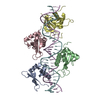
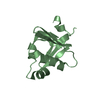
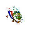
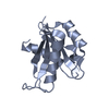
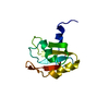
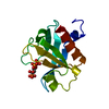
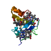
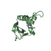
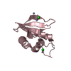
Crystal structure of IRF-3 DBD free form
Components
IRF3 protein
Keywords
DNA BINDING PROTEIN / Helix-turn-helix / Gene regulation
Function / homology
Function and homology information
IRF3 mediated activation of type 1 IFN / MDA-5 signaling pathway / macrophage apoptotic process / programmed necrotic cell death / TRIF-dependent toll-like receptor signaling pathway / LRR FLII-interacting protein 1 (LRRFIP1) activates type I IFN production / positive regulation of type I interferon-mediated signaling pathway / positive regulation of cytokine production involved in inflammatory response / IRF3-mediated induction of type I IFN / mRNA transcription ...IRF3 mediated activation of type 1 IFN / MDA-5 signaling pathway / macrophage apoptotic process / programmed necrotic cell death / TRIF-dependent toll-like receptor signaling pathway / LRR FLII-interacting protein 1 (LRRFIP1) activates type I IFN production / positive regulation of type I interferon-mediated signaling pathway / positive regulation of cytokine production involved in inflammatory response / IRF3-mediated induction of type I IFN / mRNA transcription / TRAF6 mediated IRF7 activation / cGAS/STING signaling pathway / toll-like receptor 4 signaling pathway / DNA-binding transcription activator activity / type I interferon-mediated signaling pathway / cellular response to exogenous dsRNA / cytoplasmic pattern recognition receptor signaling pathway / positive regulation of interferon-alpha production / immune system process / positive regulation of type I interferon production / positive regulation of interferon-beta production / lipopolysaccharide-mediated signaling pathway / TICAM1-dependent activation of IRF3/IRF7 / antiviral innate immune response / Regulation of innate immune responses to cytosolic DNA / Activation of IRF3, IRF7 mediated by TBK1, IKKε (IKBKE) / Negative regulators of DDX58/IFIH1 signaling / promoter-specific chromatin binding / cellular response to virus / DDX58/IFIH1-mediated induction of interferon-alpha/beta / DNA-binding transcription repressor activity, RNA polymerase II-specific / Evasion by RSV of host interferon responses / ISG15 antiviral mechanism / Interferon gamma signaling / sequence-specific double-stranded DNA binding / SARS-CoV-1 activates/modulates innate immune responses / Interferon alpha/beta signaling / regulation of inflammatory response / TRAF3-dependent IRF activation pathway / DNA-binding transcription activator activity, RNA polymerase II-specific / regulation of apoptotic process / sequence-specific DNA binding / defense response to virus / DNA-binding transcription factor activity, RNA polymerase II-specific / positive regulation of canonical NF-kappaB signal transduction / RNA polymerase II cis-regulatory region sequence-specific DNA binding / DNA-binding transcription factor activity / protein domain specific binding / apoptotic process / DNA damage response / regulation of transcription by RNA polymerase II / chromatin / SARS-CoV-2 activates/modulates innate and adaptive immune responses / protein homodimerization activity / positive regulation of transcription by RNA polymerase II / mitochondrion / DNA binding / nucleoplasm / identical protein binding / nucleus / cytoplasm / cytosol Similarity search - Function
In the structure databanks used in Yorodumi, some data are registered as the other names, "COVID-19 virus" and "2019-nCoV". Here are the details of the virus and the list of structure data.
Jan 31, 2019. EMDB accession codes are about to change! (news from PDBe EMDB page)
EMDB accession codes are about to change! (news from PDBe EMDB page)
The allocation of 4 digits for EMDB accession codes will soon come to an end. Whilst these codes will remain in use, new EMDB accession codes will include an additional digit and will expand incrementally as the available range of codes is exhausted. The current 4-digit format prefixed with “EMD-” (i.e. EMD-XXXX) will advance to a 5-digit format (i.e. EMD-XXXXX), and so on. It is currently estimated that the 4-digit codes will be depleted around Spring 2019, at which point the 5-digit format will come into force.
The EM Navigator/Yorodumi systems omit the EMD- prefix.
Related info.:Q: What is EMD? / ID/Accession-code notation in Yorodumi/EM Navigator
Yorodumi is a browser for structure data from EMDB, PDB, SASBDB, etc.
This page is also the successor to EM Navigator detail page, and also detail information page/front-end page for Omokage search.
The word "yorodu" (or yorozu) is an old Japanese word meaning "ten thousand". "mi" (miru) is to see.
Related info.:EMDB / PDB / SASBDB / Comparison of 3 databanks / Yorodumi Search / Aug 31, 2016. New EM Navigator & Yorodumi / Yorodumi Papers / Jmol/JSmol / Function and homology information / Changes in new EM Navigator and Yorodumi
 Movie
Movie Controller


 Open data
 Basic information
 Components
 Keywords
 Function and homology information
 Homo sapiens (human)
 X-RAY DIFFRACTION /
  Authors
 Citation
 Structure visualization
 Downloads & links
 Other downloads


 PDBj
PDBj


 Assembly
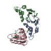


 Mass: 65.409 Da / Num. of mol.: 9 / Source method: obtained synthetically / Formula: Zn
 Mass: 35.453 Da / Num. of mol.: 7 / Source method: obtained synthetically / Formula: Cl
 Mass: 22.990 Da / Num. of mol.: 3 / Source method: obtained synthetically / Formula: Na
 Mass: 18.015 Da / Num. of mol.: 151 / Source method: isolated from a natural source / Formula: H2O
 Sample preparation
 / Beamline: 17-BM / Wavelength: 0.9798 Å
 Processing