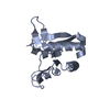
 Movie
Movie Controller


+ Open data

- Basic information
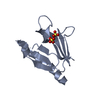
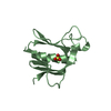
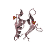
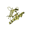
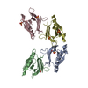
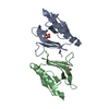
| Entry | Database: PDB / ID: 1u5d | ||||||
|---|---|---|---|---|---|---|---|
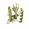
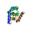
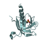
| Title | Crystal Structure of the PH domain of SKAP55 | ||||||
 Components Components | Src Kinase-associated Phosphoprotein of 55 kDa | ||||||
 Keywords Keywords | SIGNALING PROTEIN / PH Domain | ||||||
| Function / homology |  Function and homology information Function and homology informationpositive regulation of leukocyte cell-cell adhesion / positive regulation of integrin activation / positive regulation of adaptive immune response / positive regulation of cell-cell adhesion mediated by integrin / positive regulation of heterotypic cell-cell adhesion / T cell receptor complex / positive regulation of cell-matrix adhesion / immunological synapse / plasma membrane raft / SH2 domain binding ...positive regulation of leukocyte cell-cell adhesion / positive regulation of integrin activation / positive regulation of adaptive immune response / positive regulation of cell-cell adhesion mediated by integrin / positive regulation of heterotypic cell-cell adhesion / T cell receptor complex / positive regulation of cell-matrix adhesion / immunological synapse / plasma membrane raft / SH2 domain binding / protein localization to plasma membrane / SH3 domain binding / cell-cell junction / T cell receptor signaling pathway / protein phosphatase binding / adaptive immune response / protein kinase binding / positive regulation of DNA-templated transcription / positive regulation of transcription by RNA polymerase II / nucleoplasm / plasma membrane / cytoplasm / cytosol Similarity search - Function | ||||||
| Biological species |  Homo sapiens (human) Homo sapiens (human) | ||||||
| Method |  X-RAY DIFFRACTION / SYNCHROTRON / MOLECULAR REPLACEMENT / Resolution: 1.7 Å X-RAY DIFFRACTION / SYNCHROTRON / MOLECULAR REPLACEMENT / Resolution: 1.7 Å | ||||||
 Authors Authors | Tang, Y. / Swanson, K.D. / Neel, B.G. / Eck, M.J. | ||||||
 Citation Citation | Journal: To be Published Title: Structural Basis for the Dimerization and Phosphoinositide Specificity of the Src Kinase-associated Phosphoproteins SKAP55 and SKAP-Hom Authors: Tang, Y. / Swanson, K.D. / Neel, B.G. / Eck, M.J. | ||||||
| History |
|
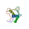
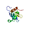
- Structure visualization
| Structure viewer | Molecule: MolmilJmol/JSmol |
|---|
- Downloads & links
-Download
| PDBx/mmCIF format | 1u5d.cif.gz | 110.7 KB | Display | PDBx/mmCIF format |
|---|---|---|---|---|
| PDB format | pdb1u5d.ent.gz | 86.1 KB | Display | PDB format |
| PDBx/mmJSON format | 1u5d.json.gz | Tree view | PDBx/mmJSON format | |
| Others |  Other downloads Other downloads |
-Validation report
| Arichive directory | https://data.pdbj.org/pub/pdb/validation_reports/u5/1u5dftp://data.pdbj.org/pub/pdb/validation_reports/u5/1u5d | HTTPS FTP |
|---|
-Related structure data
| Related structure data | 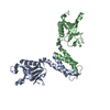 1u5eC  1u5fC  1u5gS S: Starting model for refinement C: citing same article ( |
|---|---|
| Similar structure data |


-Links
 PDBj
PDBj

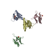
- Assembly


-Components
| #1: Protein | Mass: 12723.417 Da / Num. of mol.: 4 / Fragment: PH Domain (residues 106-213) / Mutation: D106G, N107S Source method: isolated from a genetically manipulated source Source: (gene. exp.) Homo sapiens (human) / Gene: HSSKAP55 / Plasmid: pET / Species (production host): Escherichia coli / Production host:  #2: Chemical | ChemComp-SO4 /   Mass: 96.063 Da / Num. of mol.: 7 / Source method: obtained synthetically / Formula: SO4 Mass: 96.063 Da / Num. of mol.: 7 / Source method: obtained synthetically / Formula: SO4#3: Water | ChemComp-HOH / |  Mass: 18.015 Da / Num. of mol.: 427 / Source method: isolated from a natural source / Formula: H2O Mass: 18.015 Da / Num. of mol.: 427 / Source method: isolated from a natural source / Formula: H2O |
|---|
-Experimental details
-Experiment
| Experiment | Method: X-RAY DIFFRACTION / Number of used crystals: 1 |
|---|
- Sample preparation
| Crystal | Density Matthews: 2.5 Å3/Da / Density % sol: 51.3 % |
|---|---|
| Crystal grow | Temperature: 298 K / Method: vapor diffusion, hanging drop / pH: 5 Details: Ammonium Sulfate, Sodium Acetate, pH 5.0, VAPOR DIFFUSION, HANGING DROP, temperature 298K |
-Data collection
| Diffraction | Mean temperature: 100 K |
|---|---|
| Diffraction source | Source: SYNCHROTRON / Site: APS  / Beamline: 8-BM / Wavelength: 0.978 Å / Beamline: 8-BM / Wavelength: 0.978 Å |
| Detector | Detector: CCD / Date: Dec 1, 2003 |
| Radiation | Monochromator: graphite / Protocol: SINGLE WAVELENGTH / Monochromatic (M) / Laue (L): M / Scattering type: x-ray |
| Radiation wavelength | Wavelength: 0.978 Å / Relative weight: 1 |
| Reflection | Resolution: 1.7→35 Å / Num. all: 45815 / Num. obs: 45007 / % possible obs: 83.2 % / Observed criterion σ(F): 0 / Observed criterion σ(I): 0 / Redundancy: 4.7 % / Biso Wilson estimate: 17.6 Å2 / Rmerge(I) obs: 0.04 / Net I/σ(I): 30 |
| Reflection shell | Resolution: 1.7→1.76 Å / Rmerge(I) obs: 0.183 / Mean I/σ(I) obs: 4 / Num. unique all: 2179 / % possible all: 40.3 |
- Processing
| Software |
| |||||||||||||||||||||||||
|---|---|---|---|---|---|---|---|---|---|---|---|---|---|---|---|---|---|---|---|---|---|---|---|---|---|---|
| Refinement | Method to determine structure: MOLECULAR REPLACEMENT Starting model: Structure of the PH Domain of SKAP-Hom, PDb entry 1u5g Resolution: 1.7→35 Å / Isotropic thermal model: Isotropic / Cross valid method: THROUGHOUT / σ(F): 0 / σ(I): 0 / Stereochemistry target values: MAXIMUM LIKELIHOOD
| |||||||||||||||||||||||||
| Displacement parameters | Biso mean: 24.8 Å2 | |||||||||||||||||||||||||
| Refinement step | Cycle: LAST / Resolution: 1.7→35 Å
| |||||||||||||||||||||||||
| Refine LS restraints |
| |||||||||||||||||||||||||
| LS refinement shell | Resolution: 1.7→1.74 Å
|
