 Movie
Movie Controller

[English] 日本語
 Yorodumi
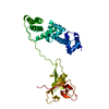
- PDB-1u5e: Crystal Structure of a N-terminal Fragment of SKAP-Hom Containing... -
+ Open data

- Basic information
| Entry | Database: PDB / ID: 1u5e | ||||||
|---|---|---|---|---|---|---|---|
| Title | Crystal Structure of a N-terminal Fragment of SKAP-Hom Containing Both the Helical Dimerization Domain and the PH Domain | ||||||
 Components Components | Src-associated adaptor protein | ||||||
 Keywords Keywords | SIGNALING PROTEIN / Novel Dimerization Domain / PH Domain | ||||||
| Function / homology |  Function and homology information Function and homology informationSignal regulatory protein family interactions / B cell activation / negative regulation of cell population proliferation / nucleoplasm / plasma membrane / cytoplasm / cytosol Similarity search - Function | ||||||
| Biological species |  | ||||||
| Method |  X-RAY DIFFRACTION / SYNCHROTRON / MOLECULAR REPLACEMENT / Resolution: 2.6 Å X-RAY DIFFRACTION / SYNCHROTRON / MOLECULAR REPLACEMENT / Resolution: 2.6 Å | ||||||
 Authors Authors | Tang, Y. / Swanson, K.D. / Neel, B.G. / Eck, M.J. | ||||||
 Citation Citation | Journal: To be Published Title: Structural Basis for the Dimerization and Phosphoinositide Specificity of the Src Kinase-associated Phosphoproteins SKAP55 and SKAP-Hom Authors: Tang, Y. / Swanson, K.D. / Neel, B.G. / Eck, M.J. | ||||||
| History |
|
- Structure visualization
| Structure viewer | Molecule: MolmilJmol/JSmol |
|---|
- Downloads & links
-Download
| PDBx/mmCIF format | 1u5e.cif.gz | 83.8 KB | Display | PDBx/mmCIF format |
|---|---|---|---|---|
| PDB format | pdb1u5e.ent.gz | 62.8 KB | Display | PDB format |
| PDBx/mmJSON format | 1u5e.json.gz | Tree view | PDBx/mmJSON format | |
| Others |  Other downloads Other downloads |
-Validation report
| Arichive directory | https://data.pdbj.org/pub/pdb/validation_reports/u5/1u5eftp://data.pdbj.org/pub/pdb/validation_reports/u5/1u5e | HTTPS FTP |
|---|
-Related structure data
| Related structure data |  1u5dC 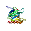 1u5fC  1u5gS S: Starting model for refinement C: citing same article ( |
|---|---|
| Similar structure data |

-Links
 PDBj
PDBj
- Assembly
| Deposited unit | 
| ||||||||
|---|---|---|---|---|---|---|---|---|---|
| 1 |
| ||||||||
| Unit cell |
| ||||||||
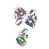
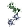
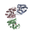
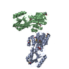
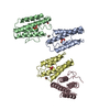
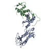
| Details | Biological dimer |
-Components
| #1: Protein | Mass: 24171.980 Da / Num. of mol.: 2 / Fragment: N-terminal Fragment, residues 12-222 / Mutation: P12G, L13S Source method: isolated from a genetically manipulated source Source: (gene. exp.)  #2: Water | ChemComp-HOH / |  Mass: 18.015 Da / Num. of mol.: 23 / Source method: isolated from a natural source / Formula: H2O Mass: 18.015 Da / Num. of mol.: 23 / Source method: isolated from a natural source / Formula: H2O |
|---|
-Experimental details
-Experiment
| Experiment | Method: X-RAY DIFFRACTION / Number of used crystals: 1 |
|---|
- Sample preparation
| Crystal | Density Matthews: 2.9 Å3/Da / Density % sol: 57.3 % |
|---|---|
| Crystal grow | Temperature: 298 K / Method: vapor diffusion, hanging drop / pH: 7.6 Details: PEG 4000, pH 7.6, VAPOR DIFFUSION, HANGING DROP, temperature 298K |
-Data collection
| Diffraction | Mean temperature: 100 K |
|---|---|
| Diffraction source | Source: SYNCHROTRON / Site: CHESS  / Beamline: A1 / Wavelength: 0.976 Å / Beamline: A1 / Wavelength: 0.976 Å |
| Detector | Detector: CCD / Date: Oct 6, 2003 / Details: mirrors |
| Radiation | Monochromator: graphite / Protocol: SINGLE WAVELENGTH / Monochromatic (M) / Laue (L): M / Scattering type: x-ray |
| Radiation wavelength | Wavelength: 0.976 Å / Relative weight: 1 |
| Reflection | Resolution: 2.6→35 Å / Num. all: 16284 / Num. obs: 15738 / % possible obs: 95.5 % / Observed criterion σ(F): 0 / Observed criterion σ(I): 0 / Redundancy: 7.8 % / Biso Wilson estimate: 58 Å2 / Rmerge(I) obs: 0.059 / Net I/σ(I): 11.9 |
| Reflection shell | Resolution: 2.6→2.66 Å / Rmerge(I) obs: 0.39 / Mean I/σ(I) obs: 2 / Num. unique all: 1075 / % possible all: 99.3 |
- Processing
| Software |
| |||||||||||||||||||||||||
|---|---|---|---|---|---|---|---|---|---|---|---|---|---|---|---|---|---|---|---|---|---|---|---|---|---|---|
| Refinement | Method to determine structure: MOLECULAR REPLACEMENT Starting model: The PH Domain of SKAP-Hom, PDB entry 1u5g Resolution: 2.6→35 Å / Isotropic thermal model: Isotropic / Cross valid method: THROUGHOUT / σ(F): 0 / σ(I): 0 / Stereochemistry target values: MAXIMUM LIKELIHOOD
| |||||||||||||||||||||||||
| Displacement parameters | Biso mean: 60 Å2 | |||||||||||||||||||||||||
| Refinement step | Cycle: LAST / Resolution: 2.6→35 Å
| |||||||||||||||||||||||||
| Refine LS restraints |
| |||||||||||||||||||||||||
| LS refinement shell | Resolution: 2.6→2.667 Å
|
