 Movie
Movie Controller

[English] 日本語
 Yorodumi
- PDB-3fhk: Crystal structure of APC1446, B.subtilis YphP disulfide isomerase -
+ Open data

- Basic information
| Entry | Database: PDB / ID: 3fhk | ||||||
|---|---|---|---|---|---|---|---|
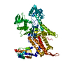
| Title | Crystal structure of APC1446, B.subtilis YphP disulfide isomerase | ||||||
 Components Components | UPF0403 protein yphP | ||||||
 Keywords Keywords | structural genomics / unknown function / disulfide isomerase / Thioredoxin superfamily / CXC motif / Surface Entropy Reduction / SER / PSI-2 / Protein structure initiative / ISFI / Integrated Center for Structure and Function Innovation | ||||||
| Function / homology |  Function and homology information Function and homology informationresponse to hydroperoxide / protein disulfide isomerase activity / cell redox homeostasis / response to oxidative stress / oxidoreductase activity Similarity search - Function | ||||||
| Biological species |  | ||||||
| Method |  X-RAY DIFFRACTION / SYNCHROTRON / MAD / Resolution: 2.3 Å X-RAY DIFFRACTION / SYNCHROTRON / MAD / Resolution: 2.3 Å | ||||||
 Authors Authors | Derewenda, U. / Boczek, T. / Cooper, D.R. / Yu, M. / Hung, L. / Derewenda, Z.S. / Integrated Center for Structure and Function Innovation (ISFI) | ||||||
 Citation Citation | Journal: Biochemistry / Year: 2009 Title: Structure and function of Bacillus subtilis YphP, a prokaryotic disulfide isomerase with a CXC catalytic motif . Authors: Derewenda, U. / Boczek, T. / Gorres, K.L. / Yu, M. / Hung, L.W. / Cooper, D. / Joachimiak, A. / Raines, R.T. / Derewenda, Z.S. | ||||||
| History |
|
- Structure visualization
| Structure viewer | Molecule: MolmilJmol/JSmol |
|---|
- Downloads & links
-Download
| PDBx/mmCIF format | 3fhk.cif.gz | 240.8 KB | Display | PDBx/mmCIF format |
|---|---|---|---|---|
| PDB format | pdb3fhk.ent.gz | 197.4 KB | Display | PDB format |
| PDBx/mmJSON format | 3fhk.json.gz | Tree view | PDBx/mmJSON format | |
| Others |  Other downloads Other downloads |
-Validation report
| Arichive directory | https://data.pdbj.org/pub/pdb/validation_reports/fh/3fhkftp://data.pdbj.org/pub/pdb/validation_reports/fh/3fhk | HTTPS FTP |
|---|
-Related structure data
| Similar structure data | |
|---|---|
| Other databases |


-Links
 PDBj
PDBj- Assembly
| Deposited unit | 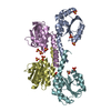
| ||||||||
|---|---|---|---|---|---|---|---|---|---|
| 1 |
| ||||||||
| 2 | 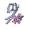
| ||||||||
| 3 | 
| ||||||||
| 4 | 
| ||||||||
| 5 | 
| ||||||||
| Unit cell |
| ||||||||
| Details | monomer |
-Components
| #1: Protein | Mass: 16049.176 Da / Num. of mol.: 4 / Mutation: Q100A, E101A Source method: isolated from a genetically manipulated source Source: (gene. exp.) #2: Chemical | ChemComp-SO4 /   Mass: 96.063 Da / Num. of mol.: 12 / Source method: obtained synthetically / Formula: SO4 Mass: 96.063 Da / Num. of mol.: 12 / Source method: obtained synthetically / Formula: SO4#3: Water | ChemComp-HOH / |  Mass: 18.015 Da / Num. of mol.: 249 / Source method: isolated from a natural source / Formula: H2O Mass: 18.015 Da / Num. of mol.: 249 / Source method: isolated from a natural source / Formula: H2O |
|---|
-Experimental details
-Experiment
| Experiment | Method: X-RAY DIFFRACTION / Number of used crystals: 1 |
|---|
- Sample preparation
| Crystal | Density Matthews: 2.73 Å3/Da / Density % sol: 54.98 % |
|---|---|
| Crystal grow | Temperature: 294 K / Method: vapor diffusion, sitting drop / pH: 6.5 Details: 2M Ammonium Sulfate, 100mM Tris, 5% PEP 629., pH 6.5, VAPOR DIFFUSION, SITTING DROP, temperature 294K |
-Data collection
| Diffraction | Mean temperature: 100 K | ||||||||||||
|---|---|---|---|---|---|---|---|---|---|---|---|---|---|
| Diffraction source | Source: SYNCHROTRON / Site: ALS  / Beamline: 8.2.2 / Wavelength: 0.97950, 0.9796, 0.9393 / Beamline: 8.2.2 / Wavelength: 0.97950, 0.9796, 0.9393 | ||||||||||||
| Detector | Type: ADSC QUANTUM 315 / Detector: CCD / Date: May 3, 2007 / Details: KOHZU: Double Crystal Si(111) | ||||||||||||
| Radiation | Protocol: MAD / Monochromatic (M) / Laue (L): M / Scattering type: x-ray | ||||||||||||
| Radiation wavelength |
| ||||||||||||
| Reflection | Resolution: 2.3→30 Å / Num. all: 32111 / Num. obs: 31840 / % possible obs: 97.2 % / Redundancy: 6.3 % / Rmerge(I) obs: 0.119 / Net I/σ(I): 19.7 | ||||||||||||
| Reflection shell | Resolution: 2.3→2.39 Å / Redundancy: 3 % / Rmerge(I) obs: 0.419 / Mean I/σ(I) obs: 2.2 / % possible all: 82.9 |
- Processing
| Software |
| ||||||||||||||||||||||||||||||||||||||||||||||||||||||||||||||||||||||||||||||||||||||||||||||||||||||||||||||||||||||||||||||||||
|---|---|---|---|---|---|---|---|---|---|---|---|---|---|---|---|---|---|---|---|---|---|---|---|---|---|---|---|---|---|---|---|---|---|---|---|---|---|---|---|---|---|---|---|---|---|---|---|---|---|---|---|---|---|---|---|---|---|---|---|---|---|---|---|---|---|---|---|---|---|---|---|---|---|---|---|---|---|---|---|---|---|---|---|---|---|---|---|---|---|---|---|---|---|---|---|---|---|---|---|---|---|---|---|---|---|---|---|---|---|---|---|---|---|---|---|---|---|---|---|---|---|---|---|---|---|---|---|---|---|---|---|
| Refinement | Method to determine structure: MAD / Resolution: 2.3→30 Å / Cor.coef. Fo:Fc: 0.95 / Cor.coef. Fo:Fc free: 0.9 / SU ML: 0.34 / Phase error: 25.7 / Stereochemistry target values: ML
| ||||||||||||||||||||||||||||||||||||||||||||||||||||||||||||||||||||||||||||||||||||||||||||||||||||||||||||||||||||||||||||||||||
| Solvent computation | Shrinkage radii: 0.9 Å / VDW probe radii: 1.11 Å / Solvent model: FLAT BULK SOLVENT MODEL / Bsol: 58.627 Å2 / ksol: 0.349 e/Å3 | ||||||||||||||||||||||||||||||||||||||||||||||||||||||||||||||||||||||||||||||||||||||||||||||||||||||||||||||||||||||||||||||||||
| Displacement parameters | Biso mean: 46.587 Å2
| ||||||||||||||||||||||||||||||||||||||||||||||||||||||||||||||||||||||||||||||||||||||||||||||||||||||||||||||||||||||||||||||||||
| Refine analyze |
| ||||||||||||||||||||||||||||||||||||||||||||||||||||||||||||||||||||||||||||||||||||||||||||||||||||||||||||||||||||||||||||||||||
| Refinement step | Cycle: LAST / Resolution: 2.3→30 Å
| ||||||||||||||||||||||||||||||||||||||||||||||||||||||||||||||||||||||||||||||||||||||||||||||||||||||||||||||||||||||||||||||||||
| Refine LS restraints |
| ||||||||||||||||||||||||||||||||||||||||||||||||||||||||||||||||||||||||||||||||||||||||||||||||||||||||||||||||||||||||||||||||||
| LS refinement shell |
| ||||||||||||||||||||||||||||||||||||||||||||||||||||||||||||||||||||||||||||||||||||||||||||||||||||||||||||||||||||||||||||||||||
| Refinement TLS params. | Refine-ID: X-RAY DIFFRACTION
| ||||||||||||||||||||||||||||||||||||||||||||||||||||||||||||||||||||||||||||||||||||||||||||||||||||||||||||||||||||||||||||||||||
| Refinement TLS group | Selection details: chain F |
