 Movie
Movie Controller


+ Open data

- Basic information
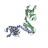
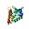
| Entry | Database: PDB / ID: 1u5g | ||||||
|---|---|---|---|---|---|---|---|
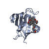
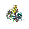
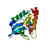
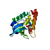
| Title | Crystal Structure of the PH Domain of SKAP-Hom | ||||||
 Components Components | Src-associated adaptor protein | ||||||
 Keywords Keywords | SIGNALING PROTEIN / PH Domain | ||||||
| Function / homology |  Function and homology information Function and homology informationSignal regulatory protein family interactions / B cell activation / negative regulation of cell population proliferation / nucleoplasm / plasma membrane / cytoplasm / cytosol Similarity search - Function | ||||||
| Biological species |  | ||||||
| Method |  X-RAY DIFFRACTION / MOLECULAR REPLACEMENT / Resolution: 2.1 Å X-RAY DIFFRACTION / MOLECULAR REPLACEMENT / Resolution: 2.1 Å | ||||||
 Authors Authors | Tang, Y. / Swanson, K. / Neel, B.G. / Eck, M.J. | ||||||
 Citation Citation | Journal: Mol.Cell / Year: 2008 Title: The Skap-hom dimerization and PH domains comprise a 3'-phosphoinositide-gated molecular switch. Authors: Swanson, K.D. / Tang, Y. / Ceccarelli, D.F. / Poy, F. / Sliwa, J.P. / Neel, B.G. / Eck, M.J. | ||||||
| History |
|
- Structure visualization
| Structure viewer | Molecule: MolmilJmol/JSmol |
|---|
- Downloads & links
-Download
| PDBx/mmCIF format | 1u5g.cif.gz | 111.5 KB | Display | PDBx/mmCIF format |
|---|---|---|---|---|
| PDB format | pdb1u5g.ent.gz | 87.2 KB | Display | PDB format |
| PDBx/mmJSON format | 1u5g.json.gz | Tree view | PDBx/mmJSON format | |
| Others |  Other downloads Other downloads |
-Validation report
| Arichive directory | https://data.pdbj.org/pub/pdb/validation_reports/u5/1u5gftp://data.pdbj.org/pub/pdb/validation_reports/u5/1u5g | HTTPS FTP |
|---|
-Related structure data
| Related structure data | 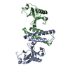 2otxC 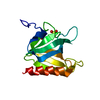 1u5fS S: Starting model for refinement C: citing same article ( |
|---|---|
| Similar structure data |


-Links
 PDBj
PDBj
- Assembly
| Deposited unit | 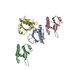
| ||||||||
|---|---|---|---|---|---|---|---|---|---|
| 1 | 
| ||||||||
| 2 | 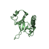
| ||||||||
| 3 | 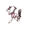
| ||||||||
| 4 | 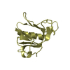
| ||||||||
| Unit cell |
|
-Components
| #1: Protein | Mass: 14208.134 Da / Num. of mol.: 4 / Fragment: PH Domain, residues 101-222 / Mutation: S101G, D102S Source method: isolated from a genetically manipulated source Source: (gene. exp.)  #2: Water | ChemComp-HOH / |  Mass: 18.015 Da / Num. of mol.: 213 / Source method: isolated from a natural source / Formula: H2O Mass: 18.015 Da / Num. of mol.: 213 / Source method: isolated from a natural source / Formula: H2O |
|---|
-Experimental details
-Experiment
| Experiment | Method: X-RAY DIFFRACTION / Number of used crystals: 1 |
|---|
- Sample preparation
| Crystal | Density Matthews: 2.7 Å3/Da / Density % sol: 54.2 % |
|---|---|
| Crystal grow | Temperature: 298 K / Method: vapor diffusion, hanging drop / pH: 7.6 Details: PEG 4000, pH 7.6, VAPOR DIFFUSION, HANGING DROP, temperature 298K |
-Data collection
| Diffraction | Mean temperature: 100 K |
|---|---|
| Diffraction source | Source: ROTATING ANODE / Type: RIGAKU / Wavelength: 1.5418 Å |
| Detector | Type: MARRESEARCH / Detector: IMAGE PLATE / Date: Jul 19, 2003 / Details: mirrors |
| Radiation | Monochromator: graphite / Protocol: SINGLE WAVELENGTH / Monochromatic (M) / Laue (L): M / Scattering type: x-ray |
| Radiation wavelength | Wavelength: 1.5418 Å / Relative weight: 1 |
| Reflection | Resolution: 2.1→25 Å / Num. all: 33609 / Num. obs: 31477 / % possible obs: 93.1 % / Observed criterion σ(F): 0 / Observed criterion σ(I): 0 / Redundancy: 7.5 % / Biso Wilson estimate: 27 Å2 / Rmerge(I) obs: 0.052 / Net I/σ(I): 16.3 |
| Reflection shell | Resolution: 2.1→2.15 Å / Rmerge(I) obs: 0.379 / Mean I/σ(I) obs: 2.9 / Num. unique all: 2178 / % possible all: 97.9 |
- Processing
| Software |
| |||||||||||||||||||||||||
|---|---|---|---|---|---|---|---|---|---|---|---|---|---|---|---|---|---|---|---|---|---|---|---|---|---|---|
| Refinement | Method to determine structure: MOLECULAR REPLACEMENT Starting model: Crystal Structure of the PH Domain of SKAP-Hom with Vector-derived N-terminal Linker Peptide, PDB entry 1U5F. Resolution: 2.1→25 Å / Isotropic thermal model: Isotropic / Cross valid method: THROUGHOUT / σ(F): 0 / σ(I): 0 / Stereochemistry target values: MAXIMUM LIKELIHOOD
| |||||||||||||||||||||||||
| Displacement parameters | Biso mean: 46 Å2 | |||||||||||||||||||||||||
| Refinement step | Cycle: LAST / Resolution: 2.1→25 Å
| |||||||||||||||||||||||||
| Refine LS restraints |
| |||||||||||||||||||||||||
| LS refinement shell | Resolution: 2.1→2.154 Å
|
