 Movie
Movie Controller


+ Open data

- Basic information
| Entry | Database: PDB / ID: 1nxj | ||||||
|---|---|---|---|---|---|---|---|
| Title | Structure of Rv3853 from Mycobacterium tuberculosis | ||||||
 Components Components | Probable S-adenosylmethionine:2-demethylmenaquinone methyltransferase | ||||||
 Keywords Keywords | UNKNOWN FUNCTION / beta/beta/alpha domain / Structural Genomics / PSI / Protein Structure Initiative / TB Structural Genomics Consortium / TBSGC | ||||||
| Function / homology |  Function and homology information Function and homology information4-hydroxy-4-methyl-2-oxoglutarate aldolase / 4-hydroxy-4-methyl-2-oxoglutarate aldolase activity / oxaloacetate decarboxylase / ribonuclease inhibitor activity / oxaloacetate decarboxylase activity / regulation of RNA metabolic process / metal ion binding Similarity search - Function | ||||||
| Biological species |   Mycobacterium tuberculosis (bacteria) Mycobacterium tuberculosis (bacteria) | ||||||
| Method |  X-RAY DIFFRACTION / SYNCHROTRON / SIRAS / Resolution: 1.9 Å X-RAY DIFFRACTION / SYNCHROTRON / SIRAS / Resolution: 1.9 Å | ||||||
 Authors Authors | Johnston, J.M. / Arcus, V.L. / Baker, E.N. / TB Structural Genomics Consortium (TBSGC) | ||||||
 Citation Citation | Journal: J.Bacteriol. / Year: 2003 Title: Crystal Structure of a Putative Methyltransferase from Mycobacterium tuberculosis: Misannotation of a Genome Clarified by Protein Structural Analysis Authors: Johnston, J.M. / Arcus, V.L. / Morton, C.J. / Parker, M.W. / Baker, E.N. | ||||||
| History |
|
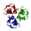
- Structure visualization
| Structure viewer | Molecule: MolmilJmol/JSmol |
|---|
- Downloads & links
-Download
| PDBx/mmCIF format | 1nxj.cif.gz | 103.9 KB | Display | PDBx/mmCIF format |
|---|---|---|---|---|
| PDB format | pdb1nxj.ent.gz | 79 KB | Display | PDB format |
| PDBx/mmJSON format | 1nxj.json.gz | Tree view | PDBx/mmJSON format | |
| Others |  Other downloads Other downloads |
-Validation report
| Arichive directory | https://data.pdbj.org/pub/pdb/validation_reports/nx/1nxjftp://data.pdbj.org/pub/pdb/validation_reports/nx/1nxj | HTTPS FTP |
|---|
-Related structure data
| Similar structure data | |
|---|---|
| Other databases |


-Links
 PDBj
PDBj
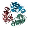
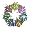
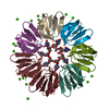
- Assembly
| Deposited unit | 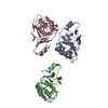
| ||||||||
|---|---|---|---|---|---|---|---|---|---|
| 1 | 
| ||||||||
| 2 | 
| ||||||||
| 3 | 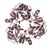
| ||||||||
| Unit cell |
| ||||||||
| Details | The biological assembly is a trimer. Trimers can be generated from each of the three monomers in the assymmetric unit by the operations: -y, x-y, z; y-x, -x, z; -x, -y, z+1/2; y, y-x, z+1/2 and x-y, x, z+1/2 |
-Components
| #1: Protein | Mass: 19380.758 Da / Num. of mol.: 3 Source method: isolated from a genetically manipulated source Source: (gene. exp.) Mycobacterium tuberculosis (bacteria) / Gene: MENG / Plasmid: pProEX Hta / Production host: References: UniProt: P0A666, UniProt: P9WGY3*PLUS, EC: 2.1.-.- #2: Chemical | 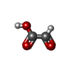  Mass: 74.035 Da / Num. of mol.: 3 / Source method: obtained synthetically / Formula: C2H2O3 Mass: 74.035 Da / Num. of mol.: 3 / Source method: obtained synthetically / Formula: C2H2O3#3: Chemical |   Mass: 150.087 Da / Num. of mol.: 3 / Source method: obtained synthetically / Formula: C4H6O6 Mass: 150.087 Da / Num. of mol.: 3 / Source method: obtained synthetically / Formula: C4H6O6#4: Water | ChemComp-HOH / |  Mass: 18.015 Da / Num. of mol.: 276 / Source method: isolated from a natural source / Formula: H2O Mass: 18.015 Da / Num. of mol.: 276 / Source method: isolated from a natural source / Formula: H2O |
|---|
-Experimental details
-Experiment
| Experiment | Method: X-RAY DIFFRACTION / Number of used crystals: 1 |
|---|
- Sample preparation
| Crystal | Density Matthews: 3.43 Å3/Da / Density % sol: 63.88 % | |||||||||||||||||||||||||||||||||||
|---|---|---|---|---|---|---|---|---|---|---|---|---|---|---|---|---|---|---|---|---|---|---|---|---|---|---|---|---|---|---|---|---|---|---|---|---|
| Crystal grow | Temperature: 291 K / Method: vapor diffusion, hanging drop / pH: 7.5 Details: 0.45M sodium potassium tartrate, pH 7.5, VAPOR DIFFUSION, HANGING DROP, temperature 291K | |||||||||||||||||||||||||||||||||||
| Crystal | *PLUS Density % sol: 63.9 % | |||||||||||||||||||||||||||||||||||
| Crystal grow | *PLUS Temperature: 18 ℃ / pH: 8 / Method: vapor diffusion, hanging drop | |||||||||||||||||||||||||||||||||||
| Components of the solutions | *PLUS
|
-Data collection
| Diffraction | Mean temperature: 110 K |
|---|---|
| Diffraction source | Source: SYNCHROTRON / Site: EMBL/DESY, HAMBURG  / Beamline: BW7B / Wavelength: 0.8452 Å / Beamline: BW7B / Wavelength: 0.8452 Å |
| Detector | Type: MARRESEARCH / Detector: IMAGE PLATE / Date: Jul 20, 2001 |
| Radiation | Protocol: SINGLE WAVELENGTH / Monochromatic (M) / Laue (L): M / Scattering type: x-ray |
| Radiation wavelength | Wavelength: 0.8452 Å / Relative weight: 1 |
| Reflection | Resolution: 1.9→25 Å / Num. all: 54630 / Num. obs: 54241 / % possible obs: 99.3 % / Redundancy: 3.3 % / Biso Wilson estimate: 9.4 Å2 / Rmerge(I) obs: 0.069 / Net I/σ(I): 14.4 |
| Reflection shell | Resolution: 1.9→1.97 Å / Rmerge(I) obs: 0.312 / Mean I/σ(I) obs: 3.1 / Num. unique all: 5249 / % possible all: 96.1 |
| Reflection | *PLUS Lowest resolution: 20 Å / Num. obs: 54630 / Redundancy: 3.35 % |
| Reflection shell | *PLUS % possible obs: 96.1 % |
- Processing
| Software |
| ||||||||||||||||||||||||||||||||||||
|---|---|---|---|---|---|---|---|---|---|---|---|---|---|---|---|---|---|---|---|---|---|---|---|---|---|---|---|---|---|---|---|---|---|---|---|---|---|
| Refinement | Method to determine structure: SIRAS / Resolution: 1.9→24.61 Å / Rfactor Rfree error: 0.003 / Isotropic thermal model: RESTRAINED / Cross valid method: THROUGHOUT / σ(F): 0 / Stereochemistry target values: Engh & Huber
| ||||||||||||||||||||||||||||||||||||
| Solvent computation | Solvent model: FLAT MODEL / Bsol: 48.1134 Å2 / ksol: 0.395904 e/Å3 | ||||||||||||||||||||||||||||||||||||
| Displacement parameters | Biso mean: 15.6 Å2
| ||||||||||||||||||||||||||||||||||||
| Refine analyze |
| ||||||||||||||||||||||||||||||||||||
| Refinement step | Cycle: LAST / Resolution: 1.9→24.61 Å
| ||||||||||||||||||||||||||||||||||||
| Refine LS restraints |
| ||||||||||||||||||||||||||||||||||||
| LS refinement shell | Resolution: 1.9→1.97 Å / Rfactor Rfree error: 0.011 / Total num. of bins used: 10
| ||||||||||||||||||||||||||||||||||||
| Xplor file |
| ||||||||||||||||||||||||||||||||||||
| Refinement | *PLUS Highest resolution: 1.9 Å / Lowest resolution: 25 Å / Rfactor Rfree: 0.22 / Rfactor Rwork: 0.19 | ||||||||||||||||||||||||||||||||||||
| Solvent computation | *PLUS | ||||||||||||||||||||||||||||||||||||
| Displacement parameters | *PLUS | ||||||||||||||||||||||||||||||||||||
| Refine LS restraints | *PLUS
| ||||||||||||||||||||||||||||||||||||
| LS refinement shell | *PLUS Num. reflection Rwork: 5344 |
