 Movie
Movie Controller

[English] 日本語
 Yorodumi
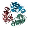
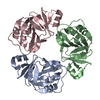
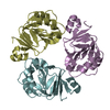
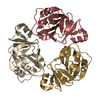
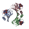
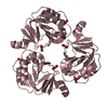
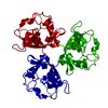
- PDB-1q5x: Structure of OF RRAA (MENG), a protein inhibitor of RNA processing -
+ Open data

- Basic information
| Entry | Database: PDB / ID: 1q5x | ||||||
|---|---|---|---|---|---|---|---|
| Title | Structure of OF RRAA (MENG), a protein inhibitor of RNA processing | ||||||
 Components Components | REGULATOR OF RNASE E ACTIVITY A | ||||||
 Keywords Keywords | hydrolase inhibitor / 3-LAYER SANDWICH / ALPHA-BETA STRUCTURE / PARALLEL BETA SHEET / ANTIPARALLEL BETA SHEET | ||||||
| Function / homology |  Function and homology information Function and homology informationnegative regulation of RNA catabolic process / ribonuclease inhibitor activity / protein homotrimerization / endoribonuclease inhibitor activity / enzyme binding / protein-containing complex / identical protein binding / cytosol Similarity search - Function | ||||||
| Biological species |  | ||||||
| Method |  X-RAY DIFFRACTION / SIRAS / Resolution: 2 Å X-RAY DIFFRACTION / SIRAS / Resolution: 2 Å | ||||||
 Authors Authors | Monzingo, A.F. / Gao, J. / Qiu, J. / Georgiou, G. / Robertus, J.D. | ||||||
 Citation Citation | Journal: J.Mol.Biol. / Year: 2003 Title: The X-ray Structure of Escherichia coli RraA (MenG), A Protein Inhibitor of RNA Processing. Authors: Monzingo, A.F. / Gao, J. / Qiu, J. / Georgiou, G. / Robertus, J.D. #1: Journal: Cell(Cambridge,Mass.) / Year: 2003Title: RraA: a protein inhibitor of RNase E activity that globally modulates RNA abundance in E. coli. Authors: LEE, K. / ZHAN, X. / GAO, J. / QIU, J. / FANG, Y. / MEGANATHAN, R. / COHEN, S.N. / GEORGIOU, G. | ||||||
| History |
| ||||||
| Remark 400 | COMPOUND THE AUTHOR MAINTAINS THAT THIS PROTEIN HAS BEEN MISANNOTATED IN THE SWISSPROT DATABASE. ...COMPOUND THE AUTHOR MAINTAINS THAT THIS PROTEIN HAS BEEN MISANNOTATED IN THE SWISSPROT DATABASE. THERE IS NO BIOCHEMICAL EVIDENCE THAT THE MENG GENE PRODUCT IS AN S-ADENOSYLMETHIONINE:2-DEMETHYLMENAQUINONE METHYLTRANSFERASE. HOWEVER, THERE IS NOW BIOCHEMICAL EVIDENCE SHOWING THAT THE PROTEIN BINDS RNASE E AND INHIBITS ITS ACTIVITY AS DISCUSSED IN REFERENCE 1. | ||||||
| Remark 700 | SHEET SHEET RECORDS IN THIS FILE WERE PROVIDED BY THE AUTHOR |
- Structure visualization
| Structure viewer | Molecule: MolmilJmol/JSmol |
|---|
- Downloads & links
-Download
| PDBx/mmCIF format | 1q5x.cif.gz | 102.6 KB | Display | PDBx/mmCIF format |
|---|---|---|---|---|
| PDB format | pdb1q5x.ent.gz | 79.9 KB | Display | PDB format |
| PDBx/mmJSON format | 1q5x.json.gz | Tree view | PDBx/mmJSON format | |
| Others |  Other downloads Other downloads |
-Validation report
| Arichive directory | https://data.pdbj.org/pub/pdb/validation_reports/q5/1q5xftp://data.pdbj.org/pub/pdb/validation_reports/q5/1q5x | HTTPS FTP |
|---|
-Related structure data
| Similar structure data |
|---|

-Links
 PDBj
PDBj- Assembly
| Deposited unit | 
| ||||||||
|---|---|---|---|---|---|---|---|---|---|
| 1 |
| ||||||||
| Unit cell |
| ||||||||
| Details | THE BIOLOGICAL UNIT IS THE HOMOTRIMER CONTAINED IN THE ASYMMETRIC UNIT |
-Components
| #1: Protein | Mass: 17371.264 Da / Num. of mol.: 3 Source method: isolated from a genetically manipulated source Source: (gene. exp.) #2: Water | ChemComp-HOH / |  Mass: 18.015 Da / Num. of mol.: 244 / Source method: isolated from a natural source / Formula: H2O Mass: 18.015 Da / Num. of mol.: 244 / Source method: isolated from a natural source / Formula: H2O |
|---|
-Experimental details
-Experiment
| Experiment | Method: X-RAY DIFFRACTION / Number of used crystals: 1 |
|---|
- Sample preparation
| Crystal | Density Matthews: 2.12 Å3/Da / Density % sol: 42.11 % | ||||||||||||||||||
|---|---|---|---|---|---|---|---|---|---|---|---|---|---|---|---|---|---|---|---|
| Crystal grow | Temperature: 298 K / Method: vapor diffusion, hanging drop / pH: 5.2 Details: AMMONIUM PHOSPHATE, SODIUM CITRATE, pH 5.2, VAPOR DIFFUSION, HANGING DROP, temperature 298K | ||||||||||||||||||
| Crystal grow | *PLUS Method: vapor diffusion, hanging drop | ||||||||||||||||||
| Components of the solutions | *PLUS
|
-Data collection
| Diffraction | Mean temperature: 103 K |
|---|---|
| Diffraction source | Source: ROTATING ANODE / Type: RIGAKU RU200 / Wavelength: 1.5418 |
| Detector | Type: RIGAKU RAXIS IV / Detector: IMAGE PLATE / Date: Jul 9, 2001 |
| Radiation | Monochromator: DOUBLE FOCUSSING MIRRORS (NI & PT) + NI FILTER Protocol: SINGLE WAVELENGTH / Monochromatic (M) / Laue (L): M / Scattering type: x-ray |
| Radiation wavelength | Wavelength: 1.5418 Å / Relative weight: 1 |
| Reflection | Resolution: 2→20 Å / Num. all: 28089 / Num. obs: 28089 / % possible obs: 95.5 % / Observed criterion σ(I): 0 / Redundancy: 2.9 % / Rmerge(I) obs: 0.071 / Net I/σ(I): 13.4 |
| Reflection shell | Resolution: 2→2.07 Å / Redundancy: 3.4 % / Rmerge(I) obs: 0.328 / % possible all: 71.8 |
| Reflection | *PLUS Lowest resolution: 20 Å / Redundancy: 2.9 % |
| Reflection shell | *PLUS % possible obs: 71.8 % / Mean I/σ(I) obs: 2.2 |
- Processing
| Software |
| ||||||||||||||||||||
|---|---|---|---|---|---|---|---|---|---|---|---|---|---|---|---|---|---|---|---|---|---|
| Refinement | Method to determine structure: SIRAS / Resolution: 2→20 Å / Cross valid method: THROUGHOUT / σ(F): 0 / Stereochemistry target values: Engh & Huber
| ||||||||||||||||||||
| Refinement step | Cycle: LAST / Resolution: 2→20 Å
| ||||||||||||||||||||
| Refine LS restraints |
| ||||||||||||||||||||
| Software | *PLUS Name: CNS / Version: 1.1 / Classification: refinement | ||||||||||||||||||||
| Refinement | *PLUS Rfactor Rfree: 0.27 / Rfactor Rwork: 0.23 | ||||||||||||||||||||
| Solvent computation | *PLUS | ||||||||||||||||||||
| Displacement parameters | *PLUS | ||||||||||||||||||||
| Refine LS restraints | *PLUS
|
