 Movie
Movie Controller

[English] 日本語
 Yorodumi
- PDB-2jlj: Crystal Structure of the cytoplasmic domain of Yersinia pestis Ys... -
+ Open data

- Basic information
| Entry | Database: PDB / ID: 2jlj | ||||||
|---|---|---|---|---|---|---|---|
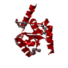
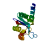
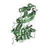
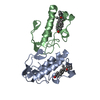
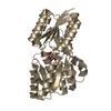
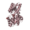
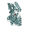
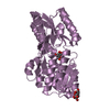
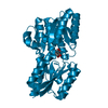
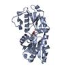
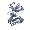
| Title | Crystal Structure of the cytoplasmic domain of Yersinia pestis YscU N263A P264A mutant | ||||||
 Components Components | YOP PROTEINS TRANSLOCATION PROTEIN U | ||||||
 Keywords Keywords | PROTEIN TRANSPORT / CELL MEMBRANE / TRANSMEMBRANE / YERSINIA PESITS / TYPE III SECRETION SYSTEM / MEMBRANE / VIRULENCE / TRANSPORT | ||||||
| Function / homology |  Function and homology information Function and homology information | ||||||
| Biological species |   YERSINIA PESTIS (bacteria) YERSINIA PESTIS (bacteria) | ||||||
| Method |  X-RAY DIFFRACTION / SYNCHROTRON / SAD / Resolution: 1.3 Å X-RAY DIFFRACTION / SYNCHROTRON / SAD / Resolution: 1.3 Å | ||||||
 Authors Authors | Lountos, G.T. / Austin, B.P. / Nallamsetty, S. / Waugh, D.S. | ||||||
 Citation Citation | Journal: Protein Sci. / Year: 2009 Title: Atomic Resolution Structure of the Cytoplasmic Domain of Yersinia Pestis Yscu, a Regulatory Switch Involved in Type III Secretion. Authors: Lountos, G.T. / Austin, B.P. / Nallamsetty, S. / Waugh, D.S. | ||||||
| History |
|
- Structure visualization
| Structure viewer | Molecule: MolmilJmol/JSmol |
|---|
- Downloads & links
-Download
| PDBx/mmCIF format | 2jlj.cif.gz | 40.9 KB | Display | PDBx/mmCIF format |
|---|---|---|---|---|
| PDB format | pdb2jlj.ent.gz | 28.3 KB | Display | PDB format |
| PDBx/mmJSON format | 2jlj.json.gz | Tree view | PDBx/mmJSON format | |
| Others |  Other downloads Other downloads |
-Validation report
| Arichive directory | https://data.pdbj.org/pub/pdb/validation_reports/jl/2jljftp://data.pdbj.org/pub/pdb/validation_reports/jl/2jlj | HTTPS FTP |
|---|
-Related structure data

-Links
 PDBj
PDBj- Assembly
| Deposited unit | 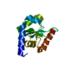
| ||||||||
|---|---|---|---|---|---|---|---|---|---|
| 1 | 
| ||||||||
| Unit cell |
| ||||||||
| Components on special symmetry positions |
|
-Components
| #1: Protein | Mass: 16881.467 Da / Num. of mol.: 1 / Fragment: CYTOPLASMIC DOMAIN, RESIDUES 211-354 / Mutation: YES Source method: isolated from a genetically manipulated source Source: (gene. exp.) YERSINIA PESTIS (bacteria) / Plasmid: PSN2006 / Production host: |
|---|---|
| #2: Water | ChemComp-HOH /  Mass: 18.015 Da / Num. of mol.: 172 / Source method: isolated from a natural source / Formula: H2O Mass: 18.015 Da / Num. of mol.: 172 / Source method: isolated from a natural source / Formula: H2O |
| Compound details | ENGINEERED |
-Experimental details
-Experiment
| Experiment | Method: X-RAY DIFFRACTION / Number of used crystals: 1 |
|---|
- Sample preparation
| Crystal | Density Matthews: 2.5 Å3/Da / Density % sol: 49.8 % / Description: NONE |
|---|---|
| Crystal grow | pH: 7 Details: 100 MM HEPES PH 7, 1 M SODIUM MALONATE PH 7, 0.75 M SODIUM CHLORIDE |
-Data collection
| Diffraction | Mean temperature: 100 K |
|---|---|
| Diffraction source | Source: SYNCHROTRON / Site: APS  / Beamline: 22-ID / Wavelength: 1 / Beamline: 22-ID / Wavelength: 1 |
| Detector | Type: MARRESEARCH / Detector: CCD / Date: Feb 5, 2008 |
| Radiation | Protocol: SINGLE WAVELENGTH / Monochromatic (M) / Laue (L): M / Scattering type: x-ray |
| Radiation wavelength | Wavelength: 1 Å / Relative weight: 1 |
| Reflection | Resolution: 1.3→50 Å / Num. obs: 37461 / % possible obs: 95.5 % / Observed criterion σ(I): 2 / Redundancy: 8.1 % / Rmerge(I) obs: 0.08 / Net I/σ(I): 19.2 |
| Reflection shell | Resolution: 1.3→1.34 Å / Redundancy: 3.1 % / Rmerge(I) obs: 0.59 / Mean I/σ(I) obs: 1.6 / % possible all: 66.5 |
- Processing
| Software |
| ||||||||||||||||||||||||||||||||||||||||||||||||||||||||||||||||||||||||||||||||||||||||||||||||||||||||||||||||||||||||||||||||||||||||||||||||||||||||||||||||||||||||||||||||||||||
|---|---|---|---|---|---|---|---|---|---|---|---|---|---|---|---|---|---|---|---|---|---|---|---|---|---|---|---|---|---|---|---|---|---|---|---|---|---|---|---|---|---|---|---|---|---|---|---|---|---|---|---|---|---|---|---|---|---|---|---|---|---|---|---|---|---|---|---|---|---|---|---|---|---|---|---|---|---|---|---|---|---|---|---|---|---|---|---|---|---|---|---|---|---|---|---|---|---|---|---|---|---|---|---|---|---|---|---|---|---|---|---|---|---|---|---|---|---|---|---|---|---|---|---|---|---|---|---|---|---|---|---|---|---|---|---|---|---|---|---|---|---|---|---|---|---|---|---|---|---|---|---|---|---|---|---|---|---|---|---|---|---|---|---|---|---|---|---|---|---|---|---|---|---|---|---|---|---|---|---|---|---|---|---|
| Refinement | Method to determine structure: SAD Starting model: NONE Resolution: 1.3→48.28 Å / Cor.coef. Fo:Fc: 0.948 / Cor.coef. Fo:Fc free: 0.932 / SU B: 1.844 / SU ML: 0.036 / TLS residual ADP flag: LIKELY RESIDUAL / Cross valid method: THROUGHOUT / ESU R: 0.056 / ESU R Free: 0.059 / Stereochemistry target values: MAXIMUM LIKELIHOOD / Details: HYDROGENS HAVE BEEN ADDED IN THE RIDING POSITIONS.
| ||||||||||||||||||||||||||||||||||||||||||||||||||||||||||||||||||||||||||||||||||||||||||||||||||||||||||||||||||||||||||||||||||||||||||||||||||||||||||||||||||||||||||||||||||||||
| Solvent computation | Ion probe radii: 0.8 Å / Shrinkage radii: 0.8 Å / VDW probe radii: 1.2 Å / Solvent model: MASK | ||||||||||||||||||||||||||||||||||||||||||||||||||||||||||||||||||||||||||||||||||||||||||||||||||||||||||||||||||||||||||||||||||||||||||||||||||||||||||||||||||||||||||||||||||||||
| Displacement parameters | Biso mean: 17.99 Å2
| ||||||||||||||||||||||||||||||||||||||||||||||||||||||||||||||||||||||||||||||||||||||||||||||||||||||||||||||||||||||||||||||||||||||||||||||||||||||||||||||||||||||||||||||||||||||
| Refinement step | Cycle: LAST / Resolution: 1.3→48.28 Å
| ||||||||||||||||||||||||||||||||||||||||||||||||||||||||||||||||||||||||||||||||||||||||||||||||||||||||||||||||||||||||||||||||||||||||||||||||||||||||||||||||||||||||||||||||||||||
| Refine LS restraints |
|