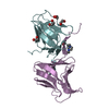
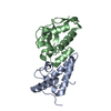
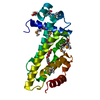
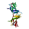
- PDB-2wkd: Crystal structure of a double Ile-to-Met mutant of protein ORF34 ... -
+
Open data
ID or keywords:
Loading...
-
Basic information
Entry
Database: PDB / ID: 2wkd
Title
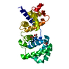
Crystal structure of a double Ile-to-Met mutant of protein ORF34 from lactococcus phage p2
Components
ORF34P2
Keywords
DNA BINDING PROTEIN / SSB / SINGLE-STRANDED DNA BINDING
Function / homology
Lactococcus phage single-stranded DNA binding protein / Lactococcus phage single-stranded DNA binding protein / Lactococcus phage SSB superfamily / Lactococcus phage single-stranded DNA binding protein / OB fold (Dihydrolipoamide Acetyltransferase, E2P) / Beta Barrel / Mainly Beta / SSB protein
Function and homology information
Biological species
LACTOCOCCUS PHAGE P2 (virus)
Method
X-RAY DIFFRACTION / SYNCHROTRON / SAD / Resolution: 2.1 Å
Mass: 18.015 Da / Num. of mol.: 26 / Source method: isolated from a natural source / Formula: H2O
Compound details
ENGINEERED RESIDUE IN CHAIN A, LEU 62 TO MSE ENGINEERED RESIDUE IN CHAIN A, LEU 68 TO MSE
Has protein modification
Y
-
Experimental details
-
Experiment
Experiment
Method: X-RAY DIFFRACTION / Number of used crystals: 1
-
Sample preparation
Crystal
Density Matthews: 4.22 Å3/Da / Density % sol: 70.9 % / Description: NONE
Crystal grow
Method: vapor diffusion, sitting drop / pH: 7.2 Details: PROTEIN AT 7 MG/ML IN 10 MM TRIS, 300 MM NACL, 1 MM TCEP, PH 8.0 WAS CRYSTALLIZED BY SITTING-DROP VAPOUR DIFFUSION AGAINST 0.1 M HEPES PH 7.2, 60% MME-PEG550
 Movie
Movie Controller

 Yorodumi
 Open data
 Basic information
 Components
 Keywords
 Function and homology information
 LACTOCOCCUS PHAGE P2 (virus)
 X-RAY DIFFRACTION /
  Authors
 Citation
 Structure visualization
 Downloads & links
 Other downloads


 PDBj
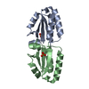
PDBj Assembly

 Mass: 18.015 Da / Num. of mol.: 26 / Source method: isolated from a natural source / Formula: H2O
 Sample preparation
 / Beamline: PROXIMA 1 / Wavelength: 0.976
  Processing